Review
. 2011 Mar;19(3):136-44.
doi: 10.1016/j.tim.2010.12.002. Epub 2010 Dec 31.

Assortment and packaging of the segmented rotavirus genome

Affiliations
Review

Assortment and packaging of the segmented rotavirus genome

Sarah M McDonald et al. Trends Microbiol. 2011 Mar.

Abstract

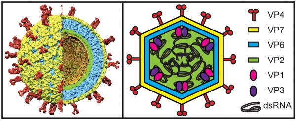
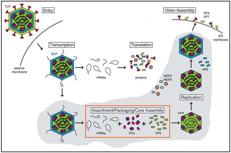
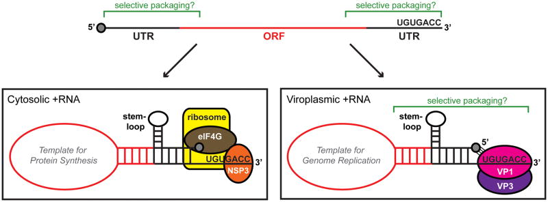
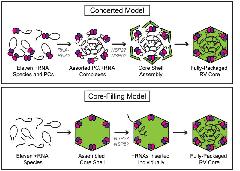
The rotavirus (RV) genome comprises 11 segments of double-stranded RNA (dsRNA) and is contained within a non-enveloped, icosahedral particle. During assembly, a highly coordinated selective packaging mechanism ensures that progeny RV virions contain one of each genome segment. Cis-acting signals thought to mediate assortment and packaging are associated with putative panhandle structures formed by base-pairing of the ends of RV plus-strand RNAs (+RNAs). Viral polymerases within assembling core particles convert the 11 distinct +RNAs to dsRNA genome segments. It remains unclear whether RV +RNAs are assorted before or during encapsidation, and the functions of viral proteins during these processes are not resolved. However, as reviewed here, recent insights gained from the study of RV and two other segmented RNA viruses, influenza A virus and bacteriophage Φ6, reveal potential mechanisms of RV assortment and packaging.


Figures

Figure 1
Figure 1. Architecture and protein composition of the RV virion
The left panel shows a cryo-electron micrograph image reconstruction of a mature, RV triple-layered particle (TLP) at 9.5 Å resolution and was used with permission from B.V.V. Prasad (Baylor University). A portion of the particle has been computationally removed to reveal the internal virion layers. The smooth external surface is made up of the VP7 glycoprotein (yellow) and is embedded with the VP4 spike attachment protein (red). The intermediate VP6 layer is shown in blue and the thin VP2 core shell is shown in green. Ordered portions of viral dsRNA that line the VP2 shell are shown in gold. Polymerase complex (PC) components, VP1 (the viral polymerase) and VP3 (the viral capping enzyme), are not visualized in this reconstruction, but are predicted to be tethered to the inner surface of VP2 near each fivefold axis. The right panel shows a cartoon schematic of a RV TLP with proteins and dsRNA colored according to the legend.
Figure 2
Figure 2. Schematic of the RV replication cycle
During entry of a RV triple-layered particle (TLP) into a host cell, VP4 and VP7 are lost, resulting in the release of a double-layered particle (DLP). The viral PCs, composed of VP1 (pink spheres) and VP3 (purple spheres), within the DLP interior are transcriptionally active and synthesize multiple copies of eleven species of capped, non-poly (A) +RNAs (black lines). The nascent +RNAs are extruded out of the DLP and deposited into the cytosol where they serve as templates for translation of viral proteins. Newly made non-structural proteins NSP2 (orange donut) and NSP5 (gray sphere) form inclusions (viroplasms; gray shaded area) around DLPs, thereby trapping +RNAs that will be used for assortment, packaging, and genome replication. Two models are proposed for how the eleven species of +RNA associate with each other, PCs, and VP2 (green) to form a fully-packaged RV core (red box; see Figure 4). During or following their encapsidation, +RNAs are used as templates for replication by the core-associated PCs to recreate eleven dsRNA segments within a pre-virion particle. A VP6 layer (blue) is acquired, and then DLPs bud into the endoplasmic reticulum (ER) during which the outer capsid proteins (VP4 and VP7) are acquired. Mature RV TLPs exit non-polarized cells predominantly by lysis.
Figure 3
Figure 3. Cis-acting elements of RV +RNAs
The top cartoon schematic represents a linear RV +RNA molecule. The central open-reading frame (ORF) is shown in red, and the 5′ and 3′ untranslated regions (UTRs) are shown in black. A cap structure (gray) is at the 5′ end of the molecule, and the consensus sequence (UGUGACC) is at the 3′ end. Regions of the +RNA thought to be important for selective packaging are indicated using green brackets. The lower left cartoon schematic represents a hypothetical cytosolic +RNA that would be used as template for protein synthesis. A panhandle structure is formed by base-pairing of the 5′ and 3′ ends, and RNA-specific stem-loop(s) are thought to project from these regions. The 3′ terminus is predicted to be bound by the nonstructural protein NSP3, which itself interacts with eukaryotic initiation factor eIF4G. The NSP3-eIF4G interaction, along with 5′-3′ complementarily, is thought to cause the +RNA to be held in a circular conformation, which might be important for efficient translation by host ribosomes. The lower right cartoon schematic represents a hypothetical viroplasmic +RNA that is selectively packaged into cores and used as a template for genome replication. Similar to the cytoplasmic +RNA, a panhandle structure is formed by base-pairing of the 5′ and 3′ ends, and RNA-specific stem-loop(s) project from these regions. The extreme 3′ end of the template is accessible to the polymerase VP1 (pink sphere) as a single-stranded tail. The 5′ cap of the template is presumed to interact with a cap-binding site on VP1. The VP3 capping enzyme is shown as a purple sphere interacting with VP1. Regions of the folded, viroplasmic +RNA thought to be important for selective packaging are indicated using green brackets.
Figure 4
Figure 4. Models of RV +RNA assortment and packaging
Two models of selective +RNA packaging, concerted or core-filling, are proposed for RV based on the strategies used by influenza A virus or Φ6, respectively. In the concerted packaging model (top), eleven species of RV +RNAs (black lines) are bound by PC components VP1 (pink spheres) and VP3 (purple spheres). These PC/+RNAs undergo assortment via gene-specific interactions among the RNA molecules. A VP2 shell (green) then assembles around the assorted PC/+RNAs to create a fully packaged, and replication-competent core. NSP2 and NSP5 may function to regulate the timing of core assembly. In the core-filling model (bottom), a VP2 shell (green) containing internally tethered PCs (pink and purple spheres), but lacking nucleic acid, first assembles. Each of the eleven +RNAs (black lines) are then individually inserted into the core, possibly by the functions of NSP2 and/or NSP5. Complete packaging triggers core expansion and initiation of genome replication. The cartoons are meant to illustrate the order of events and not the nature of protein and RNA interactions.

References

    1. Luque D, et al. Infectious bursal disease virus is an icosahedral polyploid dsRNA virus. Proc Natl Acad Sci USA. 2009;106:2148–2152. - PMC - PubMed
    1. Hundley F, et al. Genome rearrangements of bovine rotavirus after serial passage at high multiplicity of infection. Virology. 1985;143:88–103. - PubMed
    1. Joklik WK, Roner MR. What reassorts when reovirus genome segments reassort? J Bio Chem. 1995;270:4181–4184. - PubMed
    1. Patton JT. Evidence for equimolar synthesis of double-stranded RNA and minus- strand RNA in rotavirus-infected cells. Virus Res. 1990;17:199–208. - PubMed
    1. Hutchinson EC, et al. Genome packaging in influenza A virus. J Gen Virol. 2010;91:313–328. - PubMed

Publication types

MeSH terms