. 2011 Mar 1;17(5):1033-43.
doi: 10.1158/1078-0432.CCR-10-1140. Epub 2010 Dec 30.

EGFR-homing dsRNA activates cancer-targeted immune response and eliminates disseminated EGFR-overexpressing tumors in mice

Affiliations

EGFR-homing dsRNA activates cancer-targeted immune response and eliminates disseminated EGFR-overexpressing tumors in mice

Alexei Shir et al. Clin Cancer Res. .

Abstract

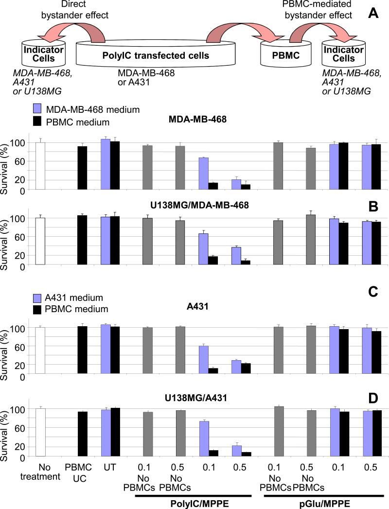
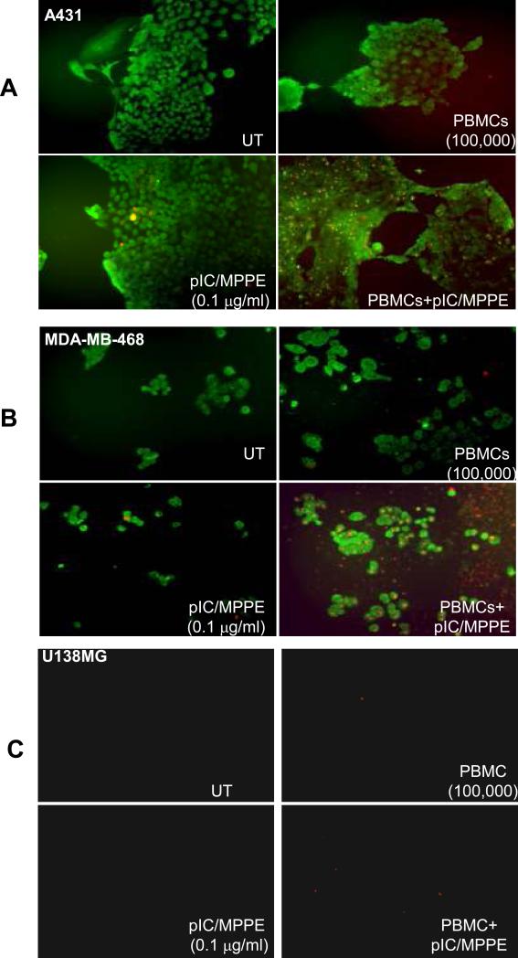
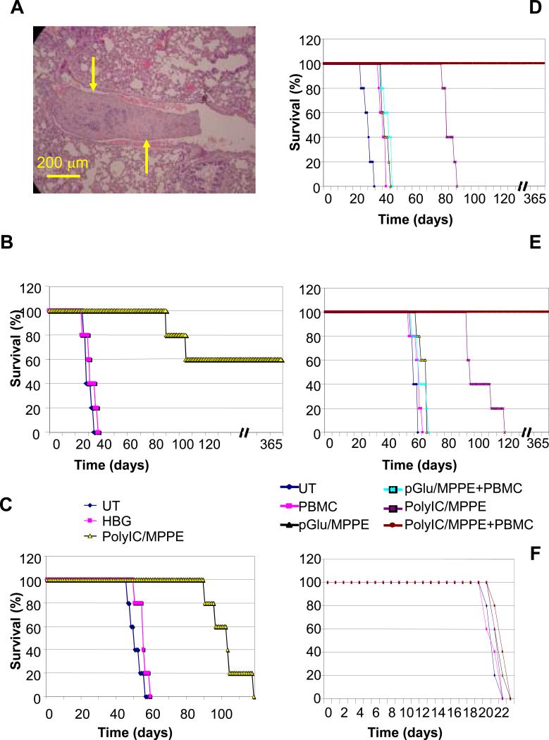
Purpose: The cause of most cancer deaths is incurable dissemination of cancer cells into vital organs. Current systemic therapies for disseminated cancers provide limited efficacy and are often accompanied by toxic side effects. We have recently shown that local application of epidermal growth factor receptor (EGFR)-targeted polyinosine-cytosine (polyIC) eradicates preestablished EGFR-overexpressing tumors. Here we show for the first time the high efficiency of systemic application of polyIC/melittin-polyethyleneimine-polyethyleneglycol-EGF (polyIC/MPPE) in combination with human immune cells.

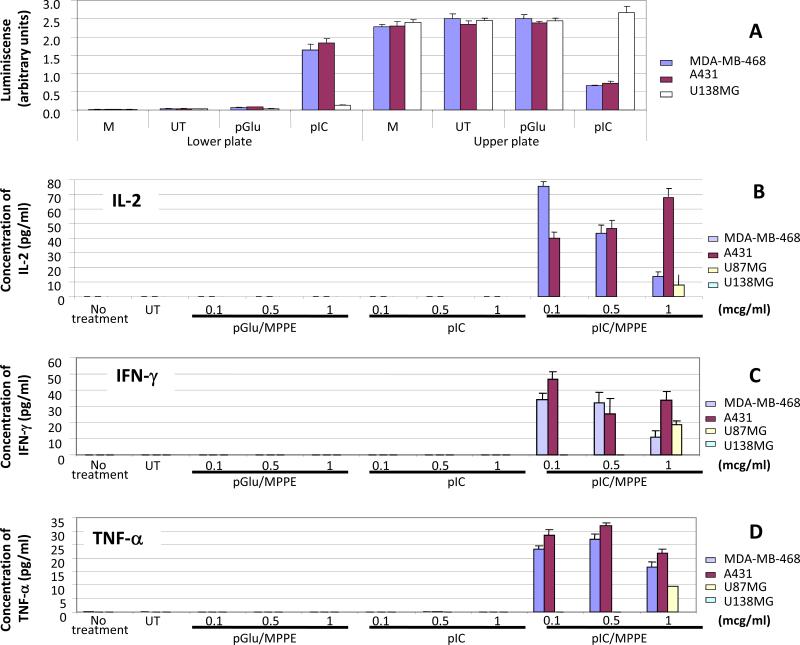
Experimental design: Cancer-targeted activation of immune cells was examined in vitro and in vivo following transfection with polyIC/MPPE. The therapeutic efficiency of the strategy was then examined on disseminated EGFR-overexpressing tumors grown in severe combined immunodeficient (SCID) mice.

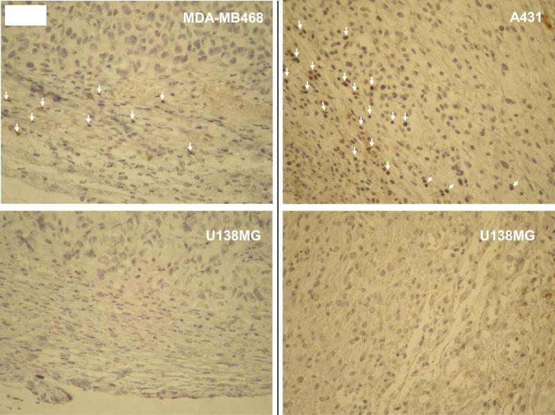
Results: Intravenous delivery of polyIC/MPPE followed by intraperitoneal injection of peripheral blood mononuclear cells induced the complete cure of SCID mice with preestablished disseminated EGFR-overexpressing tumors, with no adverse toxic effects. The immune cells and the cytokines they produce are localized to the tumor site of the treated animal and contribute decisively to the demise of the tumor cells. The immune system homes to the tumors, due to the chemokines produced by the internalized polyIC.

Conclusion: The EGFR-homing vector loaded with polyIC can be used to treat and possibly cure patients with disseminated EGFR-overexpressing tumors. The possibility of adopting this strategy to treat other tumors that express a protein capable of ligand induced internalization is discussed.


Figures

Fig. 1
Fig. 1. PBMCs are selectively attracted and activated by the medium of PolyIC/MPPE transfected MDA-MB-468 and A431 cells
(A) Chemotaxis of PBMCs. MDA-MB-468, A431 and U138MG cells were grown and treated as described in Methods. 48 hrs after the transfection medium of the cells were transferred to the lower chamber of Millipore chemotaxis plates with 5-micrometer pores. PBMCs were added to the upper chamber, and plates were incubated for 4 hrs at 37°C. Both Lower and Upper plates were then subjected to CellTiter-Glo® Luminescent Cell Viability Assay (Methods) to detect both attracted PBMCs (Lower chamber) and non-attracted PBMCs (Upper chamber). The luminescence was counted (arbitrary units) in luminometer. “M”-medium only, incubated for 48 hrs with no cells. “UT”- medium of the untreated cells. Data represent means of triplicate wells ± SD. (B-D) Activation of PBMCs. 500,000 PBMCs were seeded into 24 well plates in duplicates and grown overnight in 0.5 ml medium as described in Methods. The PBMCs were then challenged with 0.5 ml of medium removed from A431, MDA-MB-468, U87MG or U138MG cells transfected with PolyIC/PEI-PEG-EGF-Mel 48 hrs after transfection. The PBMC medium was collected 48 hours after the challenge and IFN-γ, IL-2 and TNF-α were measured using ELISA assays. Transfection with naked PolyIC as well as pGlu/MPPE (poly glutamic acid/MPPE) was used as a negative control. (B) Expression of IL-2 in the medium of PBMCs. (C) Expression of IFN–γ in the medium of PBMCs. (D) Expression of TNF-α in the medium of PBMCs. “No treatment” shows expression of the cytokines in unchallenged PBMCs. “UT” shows expression of the cytokines by PBMCs challenged with the medium of untransfected cells. Data represent means of duplicate wells ± SD, and is representative of 2 experiments.
Fig. 2
Fig. 2. Immune cells infiltrate into PolyIC/MPPE+PBMC treated EGFR over-expressing tumors
Cells were injected s.c. into the right (A431 or MDA-MB-468) and left (U138MG) flanks of SCID-NOD mice. When the tumors reached approx 100 mm3 the treatment was initiated with 3 consecutive PolyIC/MPPE I.V. injections of 20 mcg/mouse/day. 24 hrs after the last PolyIC/MPPE injection 4 million PBMCs were injected I.P. 24 hrs later tumors were extracted, and fixed in 4% formalin. Paraffin sections were then prepared, immunostained with human antiCD3 ab and subjected to histopathological analysis. White arrows indicate CD3 positive cells.
Fig. 3
Fig. 3. PBMC-mediated bystander effect
(A) Shows experiment design (described in Methods) and the bystander effect of PolyIC transfected MDA-MB-468 cells on untransfected MDA-MB-468 cells. (B) Shows the bystander effect of poly IC transfected MDA-MB-468 cells on untransfected U138MG cells. (C) Shows the bystander effect of PolyIC transfected A431 cells on untransfected A431 cells. (D) Shows the bystander effect of PolyIC transfected A431 cells on untransfected U138MG cells. “No treatment” shows survival of indicator cells that did not undergo any medium exchange. “PBMCs UC” shows survival of indicator cells treated with medium from unchallenged PBMCs. “UT” shows survival of indicator cells treated with medium from PBMCs challenged by the medium of untransfected cells. “No PBMCs” (gray bars) indicates survival when conditioned medium was added to PBMC growth medium but in the absence of PBMCs, and this was used 48 hours later to challenge the indicator cells. This latest control was used to detect a possible residual direct bystander effect of the conditioned medium after incubation in PBMCs medium in the absence of PBMCs. Mean values of the triplicates shown. The experiment was repeated twice.
Fig. 4
Fig. 4. In vitro cancer cell killing by activated PBMCs
Cells were grown as described in Methods. Cells were then transfected with PolyIC/MPPE at 0.1 mcg/ml. 24 hrs later 500,000 PBMCs/well were added to the cancer cells and co-incubated for another 24 hrs. Apoptotic cells (red fluorescence) were visualized using an Annexin-VBiotin kit (Biosource, Inc.). To distinguish tumor cells from PBMCs, tumor cells were labeled with FITC-conjugated EGFR antibody (Biosource, Inc., green fluorescence). Cells were visualized with a fluorescent microscope and photographed using a digital camera. (A) shows A431 cells, (B) shows MDA-MB-468 cells, (C) shows U138MG cells.
Fig. 5
Fig. 5. Effect of PolyIC/MPPE/PBMC treatment on survival of mice with disseminated tumors
Disseminated tumors were established as described in Methods. (A) Histopathological analysis of mouse lungs at the time of treatment initiation (15 days after cell injection). Yellow arrows point to a tumor in a lung capillary. (B, C, F) 15 days after cell injection the animals were randomly divided into groups (5 mice per group) and the treatment was initiated with 4 consecutive intravenous injections of 20 mcg PolyIC/MPPE at 24 hr intervals. 24 hrs after the last PolyIC injection, the animals were injected once with four million PBMCs. Control groups included mice treated with pGlu/MPPE (polyglutamic acid/MPPE) to determine the effect of the conjugate without PolyIC. (B) Shows survival of animals with A431 tumors. (C) Shows survival of animals with MDA-MB-468 tumors. (F) Shows survival of animals with U138MG tumors. (D, E) 10 days after cell injection the animals were randomly divided into groups (5 mice per group) and the treatment was initiated with 3 cycles of 3 or 4 consecutive intravenous injections of 20 μg polyIC/MPPE at 24 hr intervals (total 10 injections). The interval between cycles was 48 hrs. Control groups included mice treated with pGlu/MPPE (Poly Glutamic acid/MPPE) to determine the effect of the conjugate without PolyIC and HBG buffer (Hepes Buffered Glucose)(2). (D) Shows survival of animals with A431 tumors. (E) Shows survival of animals with MDA-MB-468 tumors.

References

    1. Hynes NE, MacDonald G. ErbB receptors and signaling pathways in cancer. Curr Opin Cell Biol. 2009;21:177–84. - PubMed
    1. Shir A, Ogris M, Wagner E, Levitzki A. EGF receptor-targeted synthetic double-stranded RNA eliminates glioblastoma, breast cancer, and adenocarcinoma tumors in mice. PLoS Med. 2006;3:e6. - PMC - PubMed
    1. Han Y, Caday CG, Nanda A, Cavenee WK, Huang HJ. Tyrphostin AG 1478 preferentially inhibits human glioma cells expressing truncated rather than wild-type epidermal growth factor receptors. Cancer Res. 1996;56:3859–61. - PubMed
    1. Kershaw MH, Wang G, Westwood JA, et al. Redirecting migration of T cells to chemokine secreted from tumors by genetic modification with CXCR2. Hum Gene Ther. 2002;13:1971–80. - PubMed
    1. Huang H, Xiang J. Synergistic effect of lymphotactin and interferon gamma-inducible protein-10 transgene expression in T-cell localization and adoptive T-cell therapy of tumors. Int J Cancer. 2004;109:817–25. - PubMed

Publication types

MeSH terms