. 2011 Mar;62(6):2155-68.
doi: 10.1093/jxb/erq413. Epub 2010 Dec 31.

Immunodetection of retinoblastoma-related protein and its phosphorylated form in interphase and mitotic alfalfa cells

Affiliations

Immunodetection of retinoblastoma-related protein and its phosphorylated form in interphase and mitotic alfalfa cells

Edit Abrahám et al. J Exp Bot. 2011 Mar.

Abstract

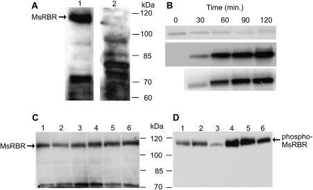
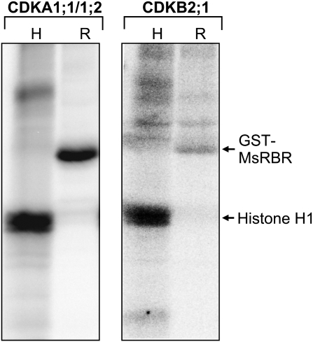
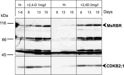
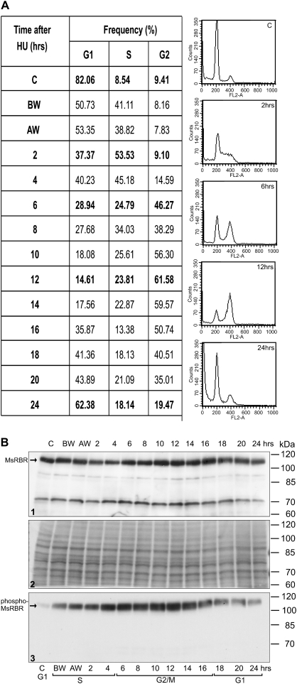
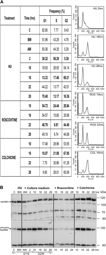
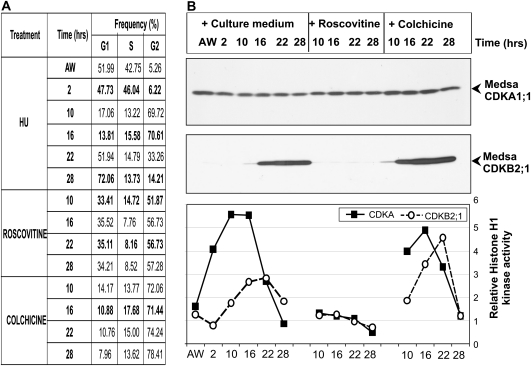
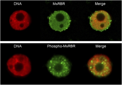
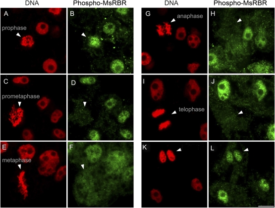
Plant retinoblastoma-related (RBR) proteins are primarily considered as key regulators of G(1)/S phase transition, with functional roles in a variety of cellular events during plant growth and organ development. Polyclonal antibody against the C-terminal region of the Arabidopsis RBR1 protein also specifically recognizes the alfalfa 115 kDa MsRBR protein, as shown by the antigen competition assay. The MsRBR protein was detected in all cell cycle phases, with a moderate increase in samples representing G(2)/M cells. Antibody against the human phospho-pRb peptide (Ser807/811) cross-reacted with the same 115 kDa MsRBR protein and with the in vitro phosphorylated MsRBR protein C-terminal fragment. Phospho-MsRBR protein was low in G(1) cells. Its amount increased upon entry into the S phase and remained high during the G(2)/M phases. Roscovitine treatment abolished the activity of alfalfa MsCDKA1;1 and MsCDKB2;1, and the phospho-MsRBR protein level was significantly decreased in the treated cells. Colchicine block increased the detected levels of both forms of MsRBR protein. Reduced levels of the MsRBR protein in cells at stationary phase or grown in hormone-free medium can be a sign of the division-dependent presence of plant RBR proteins. Immunolocalization of the phospho-MsRBR protein indicated spots of variable number and size in the labelled interphase nuclei and high signal intensity of nuclear granules in prophase. Structures similar to phospho-MsRBR proteins cannot be recognized in later mitotic phases. Based on the presented western blot and immunolocalization data, the possible involvement of RBR proteins in G(2)/M phase regulation in plant cells is discussed.


Figures

Fig. 1
Fig. 1
Specificity test of antibodies used for detection of the Medicago retinoblastoma-related protein (MsRBR) and its phosphorylated form (phospho-MsRBR) in cells at stationary or exponential growing phase. (A) Antigen competition assay for anti-AtRBR1 antibody: 1, western blot of total protein extract with anti-AtRBR1 antibody detected the MsRBR1 protein (115 kDa); 2, immunoblot of total protein extract with anti-AtRBR1 antibody pre-incubated with the purified (His)6-tagged C-terminal part of the MsRBR1 protein failed to detect the MsRBR protein (115 kDa). (B) Functionality test of anti-human pRb phosphopeptide antibody by western blot of the in vitro phosphorylated recombinant C-terminal fragment of MsRBR protein after incubation with p13SUC1-bound kinase complex. Upper panel, Ponceau S-stained filter used for immunoblot assay; middle panel, immunoblot with antibody produced against the phosphopeptide corresponding to residues around Ser807/811 of human pRb; lower panel, detection of incorporated [32P]inorganic phosphate by Phosphor Imager SI (Molecular Dynamics). (C) Alfalfa cells at exponential phase have an increased amount of the MsRBR protein in comparison with cells at stationary phase. Lanes 1–3, protein extracts from 7-day-old cultures (stationary phase); lanes 4–6, protein extracts from 4-day-old cultures (exponential phase); 1, 4, control cultures; 2, 5, protein extracts treated with phosphatase buffer; 3, 6, protein extracts with calf intestinal alkaline phosphatase (CIAP). (D) Western blots with the anti-human pRb phosphopeptide antibody detected reduced amounts of phospho-MsRBR protein after phosphatase treatment and showed significantly higher amounts of phospho-MsRBR protein in cells at exponential growing phase. Lanes 1–3, protein extracts from 7-day-old cultures (stationary phase); lanes 4–6, protein extracts from 4-day-old cultures (exponential phase); 1, 4, control cultures; 2, 5, protein extracts treated with phosphatase buffer; 3, 6, protein extracts with CIAP.
Fig. 2
Fig. 2
The MsRBR protein can serve as substrate for both the PSTAIRE (CDKA1;1/1/2) and the mitotic PPTALRE (CDKB2;1) kinases. Immunoprecipitated cyclin kinase complexes phosphorylate the histone H1 protein (H) and the MsRBR protein C-terminal fragment fused to GST (R).
Fig. 3
Fig. 3
Growth hormone-dependent presence of the MsRBR protein in cultured alfalfa cells. Western blot of total protein extracts isolated from cells grown in hormone-free medium or 2,4-dichlorophenoxy acetic acid (2,4-D)- and kinetin-supplemented medium for the indicated time periods. The anti-AtRBR1 antibody and anti-CDKB2;1 antibody detected a significant reduction in the cross-reacting proteins in cells exposed to hormone-free conditions. The low amounts of the mitotic kinase protein indicate reduced division activities in these cells. The presence of a 50 kDa protein in cells from the hormone-free treatment combination reflects the living state of the analysed cells.
Fig. 4
Fig. 4
Variation in the amounts of the MsRBR and the phospho-MsRBR proteins in alfalfa cells during cell cycle progression after synchronization with 10 mM hydroxyurea (HU) treatment. (A) Frequency data of G1, S, and G2 cells at various time points after removal of the inhibitor (HU) with characteristic DNA histograms from flow cytometry. (B) Western blot of protein extracts by anti-AtRBR1 antibody (1) and anti-human phospho-pRb peptide antibody (3). Ponceau S-stained filter with 50 μg protein samples (2). C, control, non-synchronized culture; BW, before washing out HU; AW, after washing out HU.
Fig. 5
Fig. 5
Roscovitine reduces and colchicine increases the amounts of MsRBR and phospho-MsRBR proteins in alfalfa cells exposed to double synchronization. (A) Frequency data of G1, S, and G2 cells at various time points after removal of the inhibitor [hydroxyurea (HU) at 10 mM] and treatment with roscovitine or colchicine for the indicated time periods. Characteristic DNA histograms from flow cytometry are also shown. (B) Western blot of protein extracts with anti-AtRBR1 antibody (upper) and anti-human pRb phosphopeptide antibody (low). C, control, non-synchronized culture; BW, before washing out HU; AW, after washing out HU.
Fig. 6
Fig. 6
Changes in amounts of CDKA1;1 and CDKB2;1 proteins and their activities in alfalfa cells after double synchronization based on 10 mM hydroxyurea (HU) treatment combined with roscovitine (100 μM) or colchicine (0.05%) treatment. (A) Frequency data of G1, S, and G2 cells at various time points after removal of the inhibitor (HU) subsequently grown in normal culture medium and treated with either roscovitine or colchicine. (B) Top, western blot of protein extracts with anti-CDKA1;1 antibody; middle, western blot of protein extracts with anti-CDKB2;1 antibody; bottom, histone H1 phosphorylation activities of CDKA1;1 and CDKB2;1 in the synchronized cells. AW, after washing out HU.
Fig. 7
Fig. 7
Differences in immunofluorescence labelling of MsRBR and phospho-MsRBR proteins in isolated nuclei released from alfalfa cells indicate different nuclear organization for the two forms of MsRBR protein. Upper panel: diffused, uniform staining of nucleoplasm (green) after immunolocalization with anti-AtRBR1 antibody. Lower panel: granular labelling of the nucleus (green) after immunolocalization with anti-human pRb phosphopeptide antibody. DNA stained with DAPI and pseudocoloured red.
Fig. 8
Fig. 8
Organization of the phospho-MsRBR protein into granular structures is restricted to mitotic prophase. According to the distribution of the phospho-MsRBR protein these characteristic granules cannot be detected in later mitotic stages. Immunodetection of phospho-MsRBR protein (green panels) is shown at various phases of the cell division cycle in formaldehyde-fixed suspension-cultured cells of alfalfa. Arrowheads indicate various mitotic phases (A, B, prophase; C, D, prometaphase; E, F, metaphase; G, H, anaphase; I, J, telophase) or early G1 cells (K, L). Note the significantly reduced labelling intensities at metaphase, anaphase, and telophase stages of mitosis. This figure also presents interphase nuclei (B, D, F, H, J, and L) with concentration of labelling signals in nuclear spots. Localization of phospho-MsRBR protein is shown in the green panel. DNA is stained with DAPI and pseudocoloured red (red panels). Bar=10 μm
Fig. 9
Fig. 9
Concentration of the phospho-MsRBR protein (green panel) into nuclear spots in interphase cells (A–C) and granules in prophase cells (D–I) after formaldehyde fixation. Prophase cells with advanced chromosome condensation have a considerable number of phospho-MsRBR protein-positive granules with an interchromosomal position. DNA is stained with DAPI (bar=10 μm) and pseudocoloured red (red panel).

References

    1. Ach RA, Durfee T, Miller AB, Taranto P, Hanley-Bowdoin L, Zambrski P, Gruissem W. RRB1 and RRB2 encode maize retinoblastoma-related proteins that interact with a plant D-type cyclin and geminivirus replication protein. Molecular and Cellular Biology. 1997;17:5077–5086. - PMC - PubMed
    1. Berckmans B, De Veylder L. Transcriptional control of the cell cycle. Current Opinion in Plant Biology. 2009;12:599–605. - PubMed
    1. Bhattacharyya B, Panda D, Gupta S, Banerjee M. Anti-mitotic activity of colchicine and the structural basis for its interaction with tubulin. Medicinal Research Reviews. 2008;28:155–183. - PubMed
    1. Blom N, Sicheritz-Ponten T, Gupta R, Gammeltoft S, Brunak S. Prediction of post-translational glycosylation and phosphorylation of proteins from the amino acid sequence. Proteomics. 2004;4:1633–1649. - PubMed
    1. Bögre L, Oláh Z, Dudits D. Ca2-dependent protein kinase from alfalfa (Medicago varia): partial purification and autophosphorylation. Plant Science. 1988;58:135–144.

Publication types