. 2011 Mar;52(3):518-30.
doi: 10.1194/jlr.M010603. Epub 2010 Dec 31.

Janus kinase activation by cytokine oncostatin M decreases PCSK9 expression in liver cells

Affiliations

Janus kinase activation by cytokine oncostatin M decreases PCSK9 expression in liver cells

Aiqin Cao et al. J Lipid Res. 2011 Mar.

Abstract

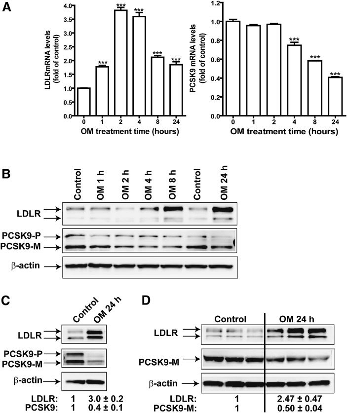
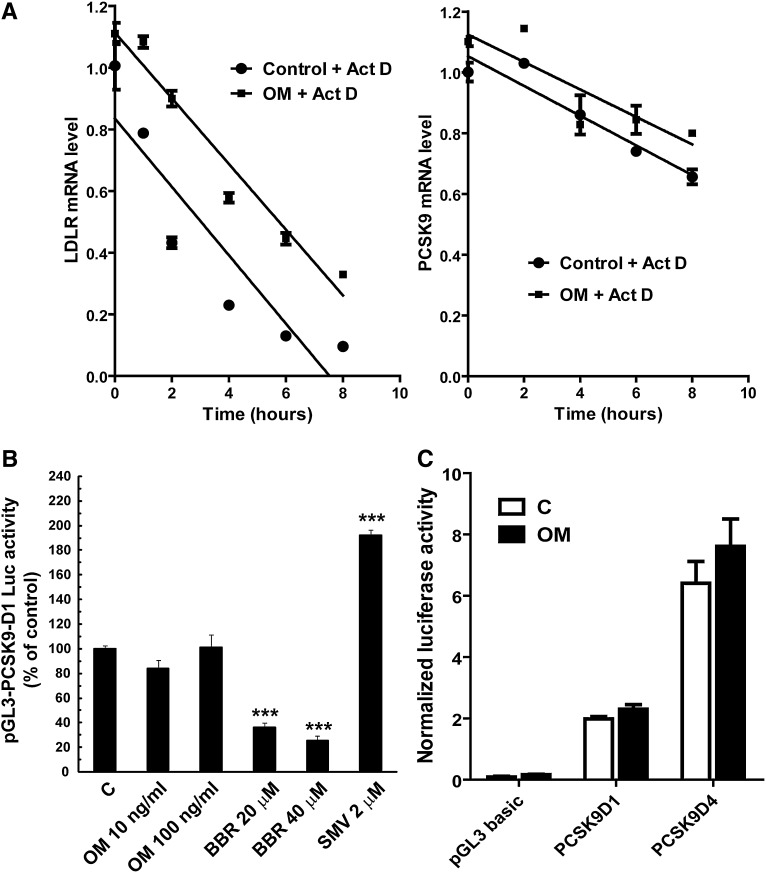
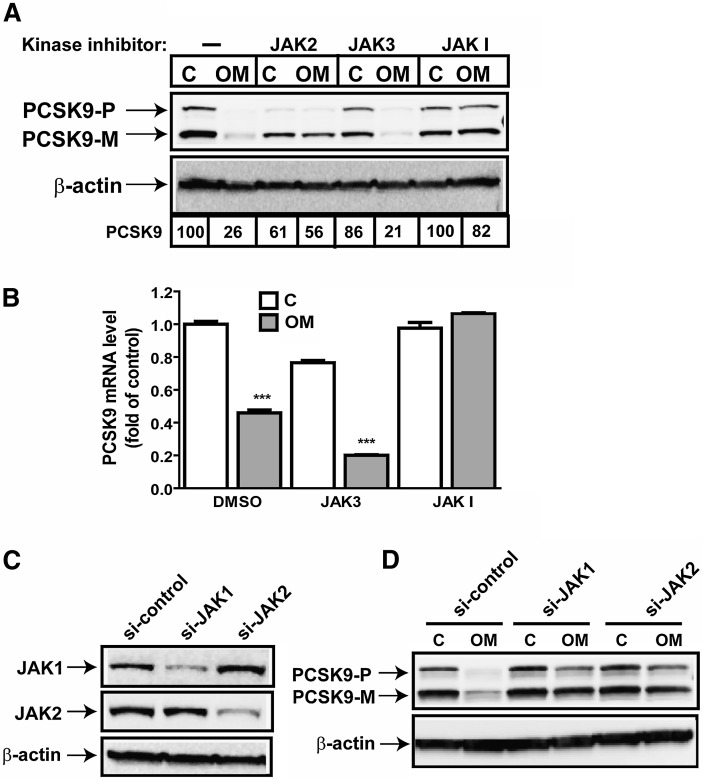
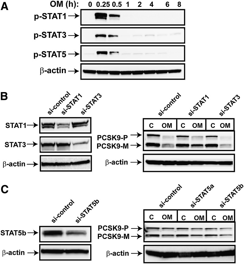
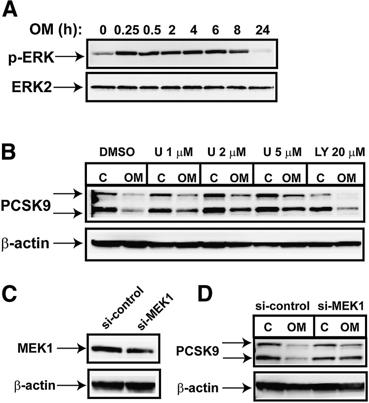
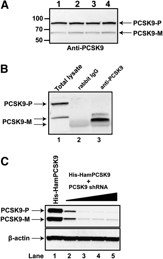
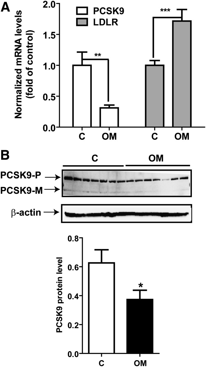
PCSK9 degrades LDL receptor (LDLR) in liver and thereby influences the circulating level of LDL cholesterol. Hence, mechanisms inhibiting PCSK9 expression have potential for cholesterol-lowering intervention. Previously, we demonstrated that oncostatin M (OM) activates LDLR gene transcription, resulting in an increased LDL uptake in HepG2 cells and a reduction of plasma LDL in hypercholesterolemic hamsters. Here we identify the suppression of PCSK9 expression by OM as one new mechanism that increases LDLR protein in HepG2 cells. Treating HepG2 cells with OM decreases PCSK9 mRNA and protein levels. Inhibition studies and small interfering RNA -targeted depletion revealed a critical role for JAK1 and JAK2 in mediating this OM inhibitory effect. Furthermore, we showed that OM induces transient phosphorylation of STAT1, STAT3, and STAT5 and sustained activation of ERK signaling molecules. While depletion of STAT members in HepG2 cells did not affect OM inhibitory activity on PCSK9 expression, blocking activation of the MEK1/ERK signaling pathway resulted in attenuation of the OM inhibitory effect. Finally, by using an anti-hamster PCSK9 antibody, we demonstrated the in vivo suppression of liver PCSK9 mRNA and protein expression by OM in hypercholesterolemic hamsters. Our study uncovered a cytokine-triggered regulatory network for PCSK9 expression that is linked to JAKs and the ERK signaling pathway.


Figures

Fig. 1.
Fig. 1.
Time-dependent effects of OM on PCSK9 mRNA and protein expression in HepG2 cells. HepG2 cells were treated with 100 ng/ml OM for different times. A: Total RNA was isolated, and mRNA levels of LDLR and PCSK9 were quantified by quantitative real-time PCR. B: Total cell lysates were isolated from HepG2 cells treated with OM from 0–24 h, and the protein abundance of LDLR and PCSK9 was examined by Western blotting. PCSK9-P, PCSK9 proprotein; PCSK9-M, mature protein. C: Cells were treated with OM for 24 h and harvested for detection of LDLR and PCSK9. The LDLR and PCSK9 bands were quantified using an Kodak Image Station 4000R. Values were normalized to β-actin and graphed relative to those of untreated cells. Data are presented as means ± SEM from six separate experiments. D: HepG2 cells were untreated or treated with OM in triplicate. The total cell lysate and medium were harvested to detect LDLR protein and PCSK9.
Fig. 2.
Fig. 2.
Examination of the OM effects on PCSK9 mRNA stability and promoter activity. A: HepG2 cells were untreated or treated with OM for 15 h. Actinomycin D (Act D) was added to cells at different intervals. Total RNA was isolated and analyzed for the amount of PCSK9 mRNA (right panel) and LDLR mRNA (left panel) by real-time PCR. Normalized PCSK9 or LDLR mRNA levels were plotted as the percentage of the mRNA remaining. Decay versus time curves were plotted. B: HepG2-derived CL26 cells were incubated with OM (100 ng/ml), BBR (20 and 40 μM), or with 2 μM of SMV for 24 h. Luciferase activities are expressed relative to that of untreated control cells. Significant differences between control and treatment groups were assessed by one-way ANOVA with a Bonferroni posttest of multiple comparisons. ***, P < 0.001 compared with untreated control cells. Data shown are representative of three separate experiments with similar results. C: HepG2 cells were transiently transfected with pGL3-PCSK9-D4 and pRL-SV40 for 1 day, and cells were treated with OM for 24 h before being lysed for dual luciferase assays.
Fig. 3.
Fig. 3.
OM-mediated suppression of PCSK9 expression requires JAK activation. A: HepG2 cells were preincubated for 2 h with JAK2 inhibitor (50 µM), JAK3 inhibitor (100 µM), or JAK inhibitor I (10 µM) prior to OM treatment for 24 h. Total cell lysates were harvested for Western blotting to detect PCSK9. PCSK9-P, PCSK9 proprotein; PCSK9-M, mature protein; C, control. B: HepG2 cells were preincubated for 2 h with JAK3 inhibitor (100 µM) or JAK inhibitor I (10 µM) prior to OM treatment for 24 h. Total RNA was prepared for real-time PCR analysis to quantify PCSK9 mRNA and GAPDH mRNA. C, D: HepG2 cells were transfected with si-JAK1, si-JAK2, or a control siRNA with a scrambled sequence for 36 h, followed by OM treatment for 24 h.
Fig. 4.
Fig. 4.
Examination of the involvement of STAT activation in OM-mediated suppression of PCSK9 protein expression. A: HepG2 cells were treated with OM at different intervals. Total cell lysates were prepared and probed for p-STAT1, p-STAT3, and p-STAT5. B, C: HepG2 cells were transfected with si-STAT1, si-STAT3, si-STAT5a, or si-STAT5b or a control (C) siRNA for 36 h, and then treated with OM for 24 h. PCSK9-P, PCSK9 proprotein; PCSK9-M, mature protein. Depletion of STAT1, STAT3, and STAT5b were detected by Western blotting with specific antibodies. The data shown are representatives of three separate transfection experiments with similar results.
Fig. 5.
Fig. 5.
MEK1/ERK signaling pathway is involved in mediating the OM effect on PCSK9 expression. A: HepG2 cells were treated with OM for different times, and total cell lysates were used to probe p-ERK1 and p-ERK2. Anti-ERK2 antibody was used to probe the total ERK in samples for protein loading control (C). B: HepG2 cells were pretreated for 2 h with inhibitor U0126 at the indicated concentrations or with PI-3 kinase inhibitor LY294002 at 20 µM for 2 h before OM was added. PCSK9-P, PCSK9 proprotein; PCSK9-M, mature protein. Cell lysates were harvested 24 h later for Western blotting of PCSK9. C, D: Cells were transfected with si-MEK1 or a control siRNA for 36 h before treatment with OM for 24 h. The protein amounts of MEK1 (C) and PCSK9 (D) were detected by Western blotting.
Fig. 6.
Fig. 6.
Comparison of hamster, mouse, rat, human, and chimpanzee PCSK9 protein sequences. The amino acid residues of mouse, rat, human, and chimpanzee PCSK9 that differed from those of the hamster sequence are boxed. Sequences with a single underline are the consensus cleavage sites. Antigen peptide residues are indicated by a double underline.
Fig. 7.
Fig. 7.
Detection of hamster PCSK9 in liver and plasma. A: Tissue protein extracts prepared from four individual hamster livers were probed with a rabbit polyclonal anti-PCSK9 antibody. Protein markers shown on the left indicate the protein molecular mass. PCSK9-P, PCSK9 proprotein; PCSK9-M, mature protein. B: A hamster serum sample was analyzed via PCSK9 immunoprecipitation and Western blotting. Total cell lysates prepared from hamster primary hepatocytes were analyzed in lane 1, the immunocomplex of rabbit normal IgG was run in lane 2, and the immunocomplex of anti-PCSK9 antibody was run in lane 3. C: HEK 293 cells were transiently transfected with pHis-HamPCSK9 alone (lane 1) or cotransfected with pSH-HamPCSK9 at the plasmid ratio of 1:1 (lane 2), 1:5 (lane 3), 1:10 (lane 4), or 1:20 (lane 5). Plasmid DNA of the empty cloning vector was used to normalize the total amount of transfected DNAs for each condition. Cell lysates were prepared 48 h after transfection, and the hamster PCSK9 protein was detected by Western blotting using the anti-hamster PCSK9 antibody.
Fig. 8.
Fig. 8.
Inhibition of PCSK9 mRNA and protein expression in liver cells of hypercholesterolemic hamsters. A: Total RNA was isolated from 50 mg of liver tissue from hamsters that were untreated or treated with 0.2 mg/kg OM for 8 days (29). Individual levels of LDLR and PCSK9 mRNA were assessed by quantitative real-time PCR using hamster-specific primers. Results are means ± SEM from 5–7 animals per group. **, P < 0.01; and ***, P < 0.001, compared with the vehicle control (C) group. B: Seven individual liver protein extracts from the control group and the OM-treated group were analyzed for PCSK9 protein expression by Western blotting. The PCSK9 protein signals were normalized to the signal intensities of β-actin individually. Values are means ± SEM. *, P < 0.05 compared with the control group. PCSK9-P, PCSK9 proprotein; PCSK9-M, mature protein.

References

    1. Seidah N. G., Benjannet S., Wickham L., Marcinkiewicz J., Bélanger J., Stifani S., Basak A., Prat A., Chrétien M. 2003. The secretory proprotein convertase neutal apoptosis-regulated convertase 1 (NARC-1): liver regeneration and neuronal differentiation. Proc. Natl. Acad. Sci. U S A. 100: 928–933. - PMC - PubMed
    1. Abifadel M., Varret M., Rabes J. P., Allard D., Ouguerram K., Devillers M., Cruaud C., Benjannet S., Wickham L., Erlich D., et al. 2003. Mutations in PCSK9 cause autosomal dominant hypercholesterolemia. Nat. Genet. 34: 154–156. - PubMed
    1. Maxwell K. N., Soccio R. E., Duncan E. M., Sehayek E., Breslow J. L. 2003. Novel putative SREBP and LXR target genes identified by microarray analysis in liver of cholesterol-fed mice. J. Lipid Res. 44: 2109–2119. - PubMed
    1. Horton J. D., Shah N. A., Warrington J. A., Anderson N. N., Park S. W., Brown M. S., Goldstein J. L. 2003. Combined analysis of oligonucleotide microarray data from transgenic and knockout mice identifies direct SREBP target genes. Proc. Natl. Acad. Sci. U S A. 100: 12027–12032. - PMC - PubMed
    1. Benjannet S., Rhainds D., Essalmani R., Mayne J., Wickham L., Jin W., Asselin M. C., Hamelin J., Varret M., Allard D., et al. 2004. NARC-1/PCSK9 and its natural mutants: zymogen cleavage and effects on the low density lipoprotein (LDL) receptor and LDL cholesterol. J. Biol. Chem. 279: 48865–48875. - PubMed

Publication types

MeSH terms