The combined effect of parathyroid hormone and bone graft on implant fixation
- PMID: 21196558
- PMCID: PMC3681808
- DOI: 10.1302/0301-620X.93B1.24261
The combined effect of parathyroid hormone and bone graft on implant fixation
Abstract
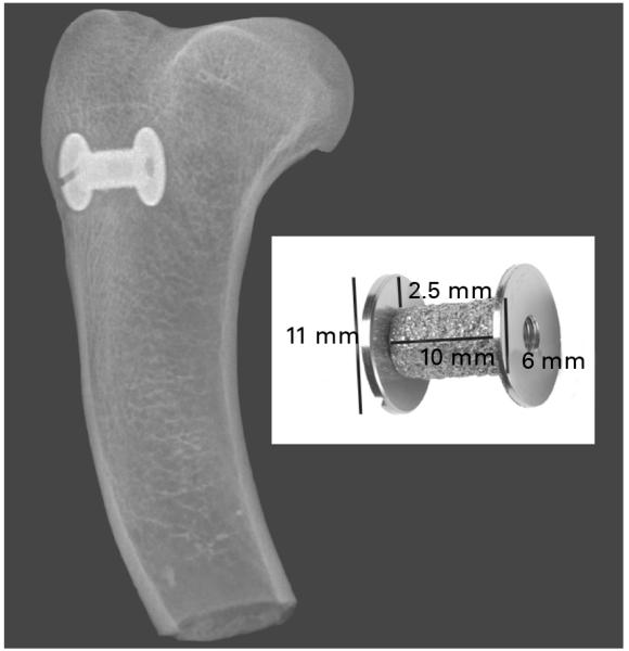
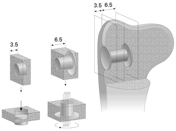
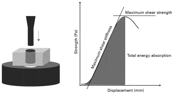
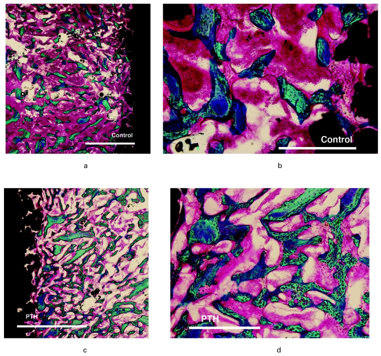
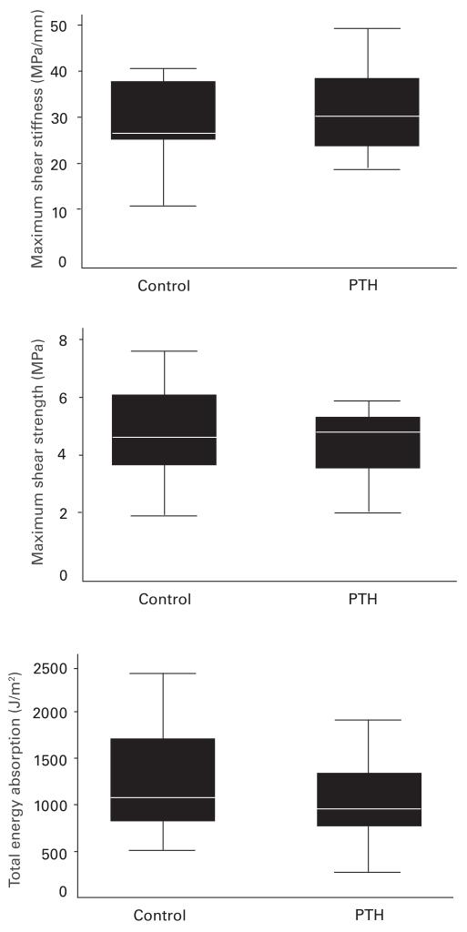
Impaction allograft is an established method of securing initial stability of an implant in arthroplasty. Subsequent bone integration can be prolonged, and the volume of allograft may not be maintained. Intermittent administration of parathyroid hormone has an anabolic effect on bone and may therefore improve integration of an implant. Using a canine implant model we tested the hypothesis that administration of parathyroid hormone may improve osseointegration of implants surrounded by bone graft. In 20 dogs a cylindrical porous-coated titanium alloy implant was inserted into normal cancellous bone in the proximal humerus and surrounded by a circumferential gap of 2.5 mm. Morsellised allograft was impacted around the implant. Half of the animals were given daily injections of human parathyroid hormone (1-34) 5 μg/kg for four weeks and half received control injections. The two groups were compared by mechanical testing and histomorphometry. We observed a significant increase in new bone formation within the bone graft in the parathyroid hormone group. There were no significant differences in the volume of allograft, bone-implant contact or in the mechanical parameters. These findings suggest that parathyroid hormone improves new bone formation in impacted morsellised allograft around an implant and retains the graft volume without significant resorption. Fixation of the implant was neither improved nor compromised at the final follow-up of four weeks.
Figures
Similar articles
-
The influence of parathyroid hormone treatment on implant fixation.Dan Med Bull. 2011 Sep;58(9):B4317. Dan Med Bull. 2011. PMID: 21893015
-
Systemic intermittent parathyroid hormone treatment improves osseointegration of press-fit inserted implants in cancellous bone.Acta Orthop. 2012 Aug;83(4):411-9. doi: 10.3109/17453674.2012.702388. Epub 2012 Aug 10. Acta Orthop. 2012. PMID: 22880714 Free PMC article.
-
Adjuvant therapies of bone graft around non-cemented experimental orthopedic implants stereological methods and experiments in dogs.Acta Orthop Suppl. 2008 Aug;79(330):1-43. Acta Orthop Suppl. 2008. PMID: 19065776 Review.
-
Soaking morselized allograft in bisphosphonate can impair implant fixation.Clin Orthop Relat Res. 2007 Oct;463:195-201. doi: 10.1097/BLO.0b013e31813c6696. Clin Orthop Relat Res. 2007. PMID: 17621234
-
Strontium in the bone-implant interface.Dan Med Bull. 2011 May;58(5):B4286. Dan Med Bull. 2011. PMID: 21535993 Review.
Cited by
-
PTH promotes allograft integration in a calvarial bone defect.Mol Pharm. 2013 Dec 2;10(12):4462-71. doi: 10.1021/mp400292p. Epub 2013 Nov 8. Mol Pharm. 2013. PMID: 24131143 Free PMC article.
-
Effect of intermittent administration of teriparatide on the mechanical and histological changes in bone grafted with β-tricalcium phosphate using a rabbit bone defect model.Exp Ther Med. 2018 Jan;15(1):19-30. doi: 10.3892/etm.2017.5424. Epub 2017 Nov 1. Exp Ther Med. 2018. PMID: 29387179 Free PMC article.
-
Teriparatide for treating delayed union and nonunion: A systematic review.J Clin Orthop Trauma. 2020 Feb;11(Suppl 1):S107-S112. doi: 10.1016/j.jcot.2019.10.009. Epub 2019 Nov 5. J Clin Orthop Trauma. 2020. PMID: 31992929 Free PMC article. Review.
-
Teriparatide as a nonoperative treatment for tibial and femoral fracture nonunion: A case report.Medicine (Baltimore). 2017 Apr;96(16):e6571. doi: 10.1097/MD.0000000000006571. Medicine (Baltimore). 2017. PMID: 28422848 Free PMC article.
-
Do Bone Graft and Cracking of the Sclerotic Cavity Improve Fixation of Titanium and Hydroxyapatite-coated Revision Implants in an Animal Model?Clin Orthop Relat Res. 2017 Feb;475(2):442-451. doi: 10.1007/s11999-016-5022-x. Clin Orthop Relat Res. 2017. PMID: 27554268 Free PMC article.
References
-
- Kärrholm J, Borssén B, Löwenhielm G, Snorrason F. Does early micromotion of femoral stem prostheses matter?: 4-7-year stereoradiographic follow-up of 84 cemented prostheses. J Bone Joint Surg [Br] 1994;76-B:912–17. - PubMed
-
- Rubash HE, Sinha RK, Shanbhag AS, Kim SY. Pathogenesis of bone loss after total hip arthroplasty. Orthop Clin North Am. 1998;29:173–86. - PubMed
-
- Gie GA, Linder L, Ling RS, et al. Impacted cancellous allografts and cement for revision total hip arthroplasty. J Bone Joint Surg [Br] 1993;75-B:14–21. - PubMed
-
- Babis GC, Sakellariou VI, O’Connor MI, Hanssen AD, Sim FH. Proximal femoral allograft-prosthesis composites in revision hip replacement: a 12-year follow-up study. J Bone Joint Surg [Br] 2010;92-B:349–55. - PubMed
-
- Brodt MD, Swan CC, Brown TD. Mechanical behavior of human morselized cancellous bone in triaxial compression testing. J Orthop Res. 1998;16:43–9. - PubMed
Publication types
MeSH terms
Substances
Grants and funding
LinkOut - more resources
Full Text Sources
Other Literature Sources
Medical
Miscellaneous