

Doppler assessment of hepatic venous waves for predicting large varices in cirrhotic patients
- PMID: 21196651
- PMCID: PMC3099078
- DOI: 10.4103/1319-3767.74465
Doppler assessment of hepatic venous waves for predicting large varices in cirrhotic patients
Abstract
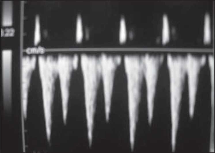
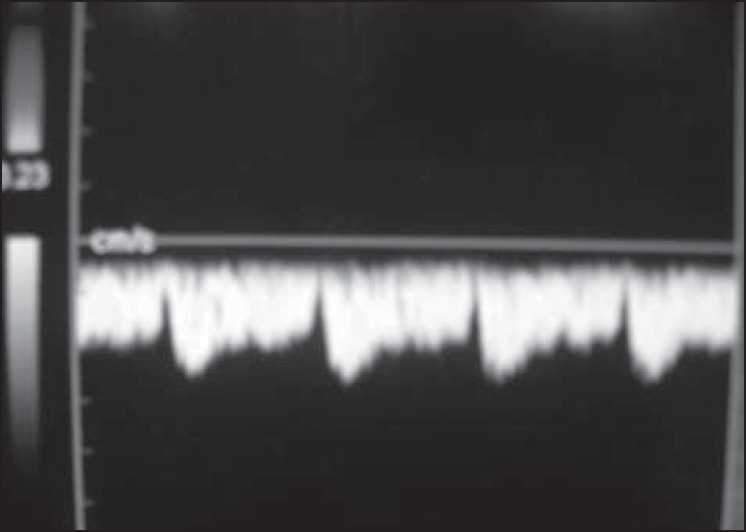
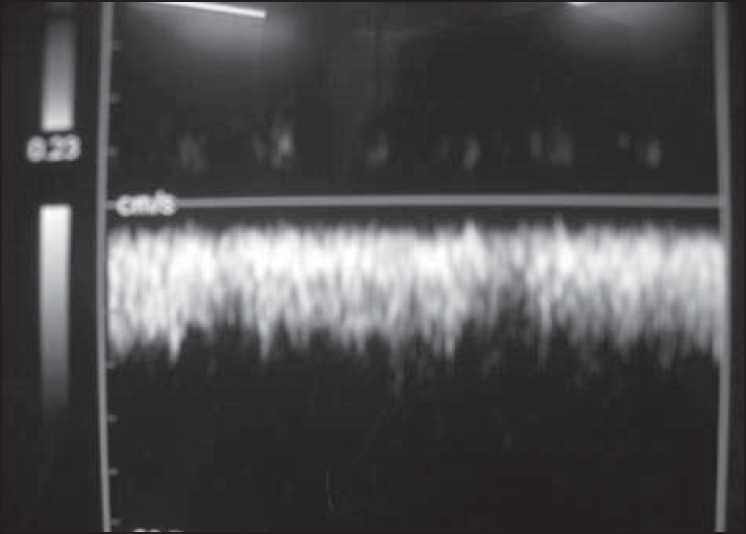
Background/aim: Color Doppler examination of changes in hepatic venous waveforms is being evaluated as a means of prediction of severity of portal hypertension and presence of esophageal varices. Normal hepatic venous waveform shows a triphasic pattern. In cirrhosis, this pattern changes to a biphasic or monophasic pattern. We aimed to study the sensitivity of loss of normal hepatic venous waveforms in predicting large varices in a cross-sectional analysis.
Materials and methods: All patients, admitted or attending the outpatient department, with a diagnosis of cirrhosis were included in the study. All patients were subjected to oesophagogastroduodenoscopy and Color Doppler examination, and waveform patterns in hepatic vein were recorded. The sensitivity and specificity of changes in waveform in detecting large varices were studied.
Results: A total of 51 cases were examined. Triphasic waves were seen in 4 (7.8%) cases, biphasic in 26 (51%) cases, and monophasic in 21 (41.2%) cases. Small varices were seen in 30 (58.8%) cases and large varices in 21 (41.2%) cases. The sensitivity of loss of the triphasic wave pattern in detecting significant varices (Grade 3 or 4) was very high (95.23%) and negative predictive value was also high (75%). Severity of liver disease as indicated by Child-Pugh and MELD scores did not correlate with changes in hepatic venous waveforms.
Conclusion: Loss of triphasic hepatic venous waveform is highly sensitive in predicting significant varices in patients with cirrhosis.
Conflict of interest statement
Figures
Similar articles
-
Hepatic vein waveform in liver cirrhosis: correlation with child's class and size of varices.J Pak Med Assoc. 2012 Aug;62(8):794-7. J Pak Med Assoc. 2012. PMID: 23862252
-
Qualitative hepatic venous Doppler sonography versus portal flowmetry in predicting the severity of esophageal varices in hepatitis C cirrhosis.AJR Am J Roentgenol. 1997 Aug;169(2):511-5. doi: 10.2214/ajr.169.2.9242766. AJR Am J Roentgenol. 1997. PMID: 9242766
-
Doppler Assessment of Hepatic Venous Waves for Evaluation of Large Varices in Cirrhotic Patient.Mymensingh Med J. 2016 Oct;25(4):641-646. Mymensingh Med J. 2016. PMID: 27941723
-
Invasive and noninvasive methods to diagnose portal hypertension and esophageal varices.Clin Liver Dis. 2014 May;18(2):293-302. doi: 10.1016/j.cld.2013.12.002. Epub 2014 Feb 25. Clin Liver Dis. 2014. PMID: 24679495 Review.
-
Evaluation and treatment of esophageal varices in the cirrhotic patient.Isr Med Assoc J. 2013 Feb;15(2):109-15. Isr Med Assoc J. 2013. PMID: 23516775 Review.
Cited by
-
Hepatic Venous Waveform, Splenoportal and Damping Index in Liver Cirrhosis: Correlation with Child Pugh's Score and Oesophageal Varices.J Clin Diagn Res. 2016 Feb;10(2):TC01-5. doi: 10.7860/JCDR/2016/15706.7181. Epub 2016 Feb 1. J Clin Diagn Res. 2016. PMID: 27042553 Free PMC article.
-
Etiological Spectrum of Cirrhosis in India: A Systematic Review and Meta-analysis.J Clin Exp Hepatol. 2024 Mar-Apr;14(2):101291. doi: 10.1016/j.jceh.2023.10.002. Epub 2023 Oct 9. J Clin Exp Hepatol. 2024. PMID: 38544766 Free PMC article.
-
Correlation Between Hepatic Waveform Changes on Doppler Ultrasound and Disease Severity in Cirrhotic Patients.Cureus. 2025 May 7;17(5):e83658. doi: 10.7759/cureus.83658. eCollection 2025 May. Cureus. 2025. PMID: 40486465 Free PMC article.
-
Prediction of Esophageal Varices in Viral Hepatitis C Cirrhosis: Performance of Combined Ultrasonography and Clinical Predictors.Int J Biomed Imaging. 2023 Sep 15;2023:7938732. doi: 10.1155/2023/7938732. eCollection 2023. Int J Biomed Imaging. 2023. PMID: 37746529 Free PMC article.
-
Hepatic Function Predictive Value of Hepatic Venous Waveform versus Portal Vein Velocity in Liver Cirrhosis.J Med Ultrasound. 2022 May 3;30(2):109-115. doi: 10.4103/JMU.JMU_91_21. eCollection 2022 Apr-Jun. J Med Ultrasound. 2022. PMID: 35832354 Free PMC article.
References
-
- Garcia-Tsao G, Sanyal AJ, Grace ND, Carey WD. Practice Guidelines Committee of the American Association for Study of Liver Diseases, Practice Parameters Committee of the American College of Gastroenterology. Prevention and management of gastroesophageal varices and variceal hemorrhage in cirrhosis. Am J Gastroenterol. 2007;102:2086–102. - PubMed
-
- Spiegel BM, Targownik L, Dulai GS, Karsan HA, Gralnek IM. Endoscopic screening for esophageal varices in cirrhosis: Is it ever cost effective? Hepatology. 2003;37:366–77. - PubMed
-
- Schepis F, Camma C, Niceforo D, Magnano A, Pallio S, Cinquegrani M, et al. Which patients with cirrhosis should undergo endoscopic screening for esophageal varices detection? Hepatology. 2001;33:333–8. - PubMed
-
- Coulden RA, Lomas DJ, Farman P, Britton PD. Doppler ultrasound of the hepatic veins: Normal appearances. Clin Radiol. 1992;45:223–7. - PubMed
-
- Abu-Yousef MM. Normal and respiratory variations of the hepatic and portal venous duplex Doppler waveforms with simultaneous electrocardiographic correlation. J Ultrasound Med. 1992;11:263–8. - PubMed
MeSH terms
LinkOut - more resources
Full Text Sources
Medical