

A case of facial deformity due to bilateral developmental maxillary cheek teeth displacement in an adult horse
- PMID: 21197210
- PMCID: PMC2942058
A case of facial deformity due to bilateral developmental maxillary cheek teeth displacement in an adult horse
Abstract
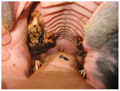
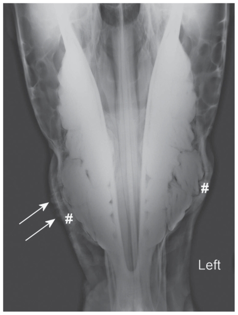
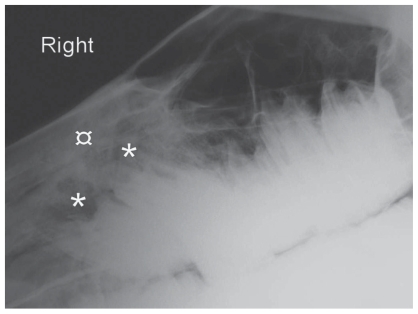
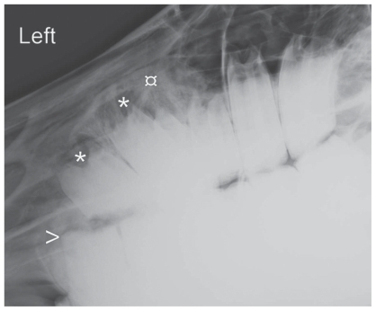
A 7-year-old mare presented with facial deformities associated with oral discomfort and weight loss was found to have bilateral, palatal, developmental displacements of the maxillary 08s, with secondary diastema. Following repulsion of both displaced teeth, the horse regained weight and resumed training. Bony deformities remained visible 9 mo after discharge.
Un cas de déformation faciale suite à un déplacement développemental bilatéral de dents molariformes chez un cheval adulte. Une jument de 7 ans présentée avec des déformations faciales associées à de l’inconfort oral et à une perte de poids a montré, après examen, des déplacements développementaux, bilatéraux, du coté palatal des 4e prémolaires supérieures, avec un diastème secondaire. Après répulsion des 2 dents déplacées, le cheval a regagné de l’état et repris l’entrainement. Les déformations osseuses étaient toujours visibles 9 mois après la sortie de la jument.
(Traduit par les auteurs)
Figures


Similar articles
-
Standing intraoral extractions of cheek teeth aided by partial crown removal in 165 horses (2010-2016).Equine Vet J. 2018 Jan;50(1):48-53. doi: 10.1111/evj.12727. Epub 2017 Aug 28. Equine Vet J. 2018. PMID: 28744895
-
Repulsion of maxillary and mandibular cheek teeth in standing horses.Vet Surg. 2011 Jul;40(5):590-5. doi: 10.1111/j.1532-950X.2011.00819.x. Epub 2011 Apr 5. Vet Surg. 2011. PMID: 21466566
-
Lateral buccotomy for removal of a supernumerary cheek tooth in a horse.J Am Vet Med Assoc. 1997 Aug 1;211(3):339-40. J Am Vet Med Assoc. 1997. PMID: 9262676
-
Equine Standing Surgical Extraction Techniques.Vet Clin North Am Equine Pract. 2020 Dec;36(3):575-612. doi: 10.1016/j.cveq.2020.08.008. Vet Clin North Am Equine Pract. 2020. PMID: 33189233 Review.
-
Adjunct Extraction Techniques in Equine Dentistry.Vet Clin North Am Equine Pract. 2020 Dec;36(3):565-574. doi: 10.1016/j.cveq.2020.08.002. Epub 2020 Oct 14. Vet Clin North Am Equine Pract. 2020. PMID: 33067099 Review.
Cited by
-
Intranasal Dental Repulsion of a Displaced Cheek Tooth in an Arabian Filly.Animals (Basel). 2025 Mar 8;15(6):772. doi: 10.3390/ani15060772. Animals (Basel). 2025. PMID: 40150301 Free PMC article.
References
-
- Floyd MR. The modified Triadan system: Nomenclature for veterinary dentistry. J Vet Dent. 1991;8:18–9. - PubMed
-
- Butler JA, Colles CM, Dyson S, Kold S, Poulos P. Clinical Radiology of the Horse. 2nd ed. Oxford: Wiley-Blackwell; 2000. pp. 327–402.
-
- Gieche JM. How to assess equine oral health. Proc Am Assoc Equine Pract. 2007;53:498–503.
-
- Dixon PM, Dacre I. A review of equine dental disorders. Vet J. 2005;169:165–187. - PubMed
-
- Easley J. Equine dental developmental abnormalities. Proc Focus Meeting on Dentistry, Am Assoc Equine Pract 2006. [Last accessed July 26, 2010]. Available from http://www.ivis.org/proceedings/aaepfocus/2006/easley2.pdf.
Publication types
MeSH terms
LinkOut - more resources
Full Text Sources
Medical