Granulosa theca cell tumor with erythrocytosis in a llama
- PMID: 21197211
- PMCID: PMC2942059
Granulosa theca cell tumor with erythrocytosis in a llama
Abstract
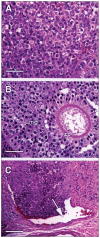
A 2.5-year-old, female llama with weight loss and lethargy had a packed cell volume (PCV) of 45% which increased to 57% over 3 wk. Transrectal ultrasonography revealed a mass of mixed echogenicity involving the right ovary, which was removed. A histopathological diagnosis of granulosa theca cell tumor was made. This is the first report of its kind in a llama.
Tumeur de la granulosa et de la thèque avec érythrocytose chez un lama. Un lama femelle âgé de 2,5 ans est présenté avec perte de poids et de la léthargie et une valeur d’hématocrite de 45 % qui a augmenté à 57 % après 3 semaines. Une échographie transrectale a révélé une masse d’échogénicité mixte touchant l’ovaire droit, qui a été retirée. Un diagnostic histopathologique d’une tumeur de la granulosa et de la thèque a été porté. Il s’agit du premier rapport de ce type chez un lama.
(Traduit par Isabelle Vallières)
Figures

References
-
- Garry F. Clinical pathology of llamas. Vet Clin N Amer: Food Animal Practice. Llama Medicine. 1989;5:55–70. - PubMed
-
- Grem JL. 5-Fluoropyrimidines. In: Chabner BA, Longo DL, editors. Cancer Chemotherapy and Biotherapy: Principles and Practice. 2nd ed. Philadelphia: Lippincott-Raven; 1996. pp. 149–211.
-
- Couto G. Tumors in Camelids: The Ohio State University Experience. Proceedings, Camelid Medicine, Surgery, and Reproduction for Veterinarians Conference; Columbus, Ohio. 2002. pp. 175–182.
-
- Hatipoglu F, Kiran MM, Ortatatli M, et al. An abattoir study of genital pathology in cows: I. Ovary and oviduct. Rev Med Vet. 2002;153:29–33.
-
- Hostetler DE, Sprecher DJ, Yamini B, Ames NK. Diagnosis and management of a malignant granulose cell tumor in a Holstein nulligravida: A case study. Theriogenology. 1997;48:11–17. - PubMed
Publication types
MeSH terms
Supplementary concepts
LinkOut - more resources
Full Text Sources
Medical
