

Development, characterization and clinical use of a biodegradable composite scaffold for bone engineering in oro-maxillo-facial surgery
- PMID: 21197218
- PMCID: PMC2946048
- DOI: 10.4161/org.6.3.12392
Development, characterization and clinical use of a biodegradable composite scaffold for bone engineering in oro-maxillo-facial surgery
Abstract
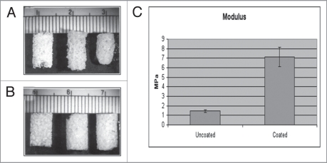
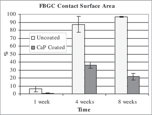
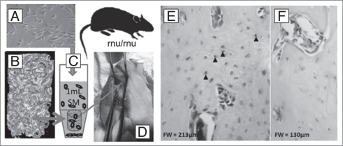
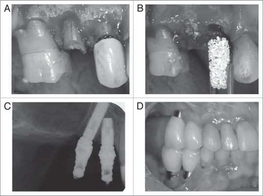
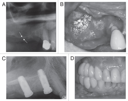
We have developed a biodegradable composite scaffold for bone tissue engineering applications with a pore size and interconnecting macroporosity similar to those of human trabecular bone. The scaffold is fabricated by a process of particle leaching and phase inversion from poly(lactideco-glycolide) (PLGA) and two calcium phosphate (CaP) phases both of which are resorbable by osteoclasts; the first a particulate within the polymer structure and the second a thin ubiquitous coating. The 3-5 μm thick osteoconductive surface CaP abrogates the putative foreign body giant cell response to the underlying polymer, while the internal CaP phase provides dimensional stability in an otherwise highly compliant structure. The scaffold may be used as a biomaterial alone, as a carrier for cells or a three-phase drug delivery device. Due to the highly interconnected macroporosity ranging from 81% to 91%, with macropores of 0.8∼1.8 mm, and an ability to wick up blood, the scaffold acts as both a clot-retention device and an osteoconductive support for host bone growth. As a cell delivery vehicle, the scaffold can be first seeded with human mesenchymal cells which can then contribute to bone formation in orthotopic implantation sites, as we show in immune-compromised animal hosts. We have also employed this scaffold in both lithomorph and particulate forms in human patients to maintain alveolar bone height following tooth extraction, and augment alveolar bone height through standard sinus lift approaches. We provide a clinical case report of both of these applications; and we show that the scaffold served to regenerate sufficient bone tissue in the wound site to provide a sound foundation for dental implant placement. At the time of writing, such implants have been in occlusal function for periods of up to 3 years in sites regenerated through the use of the scaffold.
Keywords: biodegradable; bone regeneration; cell delivery; clinical; clot retention; composite; extraction socket; osteoconduction; scaffold; sinus lift.
Figures
References
-
- Holy CE, Shoichet MS, Davies JE. Engineering three-dimensional bone tissue in vitro using biodegradable scaffolds: investigating initial cell-seeding density and culture period. J Biomed Mater Res. 2000;51:376–382. - PubMed
-
- Guan L, Davies JE. Preparation and characterization of a highly macroporous biodegradable composite tissue engineering scaffold. J Biomed Mater Res A. 2004;71:480–487. - PubMed
-
- Lickorish D, Guan L, Davies JE. A three-phase, fully resorbable, polyester/calcium phosphate scaffold for bone tissue engineering: evolution of scaffold design. Biomaterials. 2007;28:1495–1502. - PubMed
-
- Piattelli M, Favero GA, Scarano A, Orsini G, Piattelli A. Bone reactions to anorganic bovine bone (Bio-Oss) used in sinus augmentation procedures: a histological long-term report of 20 cases in humans. Int J Oral Maxillofac Implants. 1999;14:835–840. - PubMed
-
- Walsh WR, Vizesi F, Michael D, Auld J, Langdown A, Oliver R, et al. Beta-TCP bone graft substitutes in a bilateral rabbit tibial defect model. Biomaterials. 2008;29:266–271. - PubMed
Publication types
MeSH terms
Substances
Grants and funding
LinkOut - more resources
Full Text Sources
Miscellaneous