

Sensory nerve conduction and nociception in the equine lower forelimb during perineural bupivacaine infusion along the palmar nerves
- PMID: 21197231
- PMCID: PMC2949344
Sensory nerve conduction and nociception in the equine lower forelimb during perineural bupivacaine infusion along the palmar nerves
Abstract
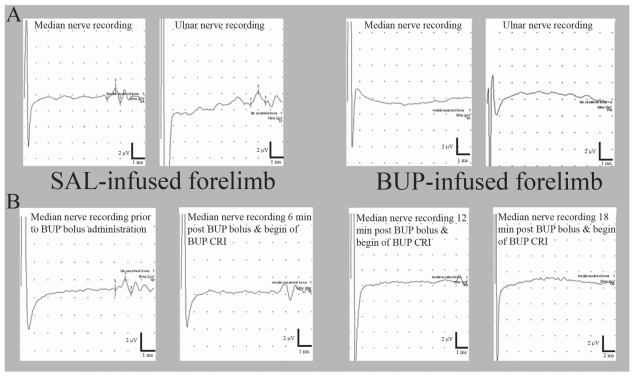
The purpose of this investigation was to study lateral palmar nerve (LPN) and medial palmar nerve (MPN) morphology and determine nociception and sensory nerve conduction velocity (SNCV) following placement of continuous peripheral nerve block (CPNB) catheters along LPN and MPN with subsequent bupivacaine (BUP) infusion. Myelinated nerve fiber distribution in LPN and MPN was examined after harvesting nerve specimens in 3 anesthetized horses and processing them for morphometric analysis. In 5 sedated horses, CPNB catheters were placed along each PN in both forelimbs. Horses then received in one forelimb 3 mL 0.125% BUP containing epinephrine 1:200 000 and 0.04% NaHCO(3) per catheter site followed by 2 mL/h infusion over a 6-day period, while in the other forelimb equal amounts of saline (SAL) solution were administered. The hoof withdrawal response (HWR) threshold during pressure loading of the area above the dorsal coronary band was determined daily in both forelimbs. On day 6 SNCV was measured under general anesthesia of horses in each limb's LPN and MPN to detect nerve injury, followed by CPNB catheter removal. The SNCV was also recorded in 2 anesthetized non-instrumented horses (sham controls). In both LPN and MPN myelinated fiber distributions were bimodal. The fraction of large fibers (>7 μm) was greater in the MPN than LPN (P < 0.05). Presence of CPNB catheters and SAL administration did neither affect measured HWR thresholds nor SNCVs, whereas BUP infusion suppressed HWRs. In conclusion, CPNB with 0.125% BUP provides pronounced analgesia by inhibiting sensory nerve conduction in the distal equine forelimb.
L’objectif de cette étude était d’étudier la morphologie du nerf palmaire latérale (LPN) et médiale (MPN) et de déterminer la nociception et la vélocité de conduction du nerf sensitif (SNCV) suite à la mise en place de cathéters pour un bloc continu du nerf périphérique (CPNB) le long de LPN et MPN avec des infusions subséquentes de buvicaïne (BUP). La distribution des fibres nerveuses myélinisées dans LPN et MPN a été examinée après la récolte de spécimens de nerf chez 3 chevaux anesthésiés et en les traitant pour analyse morphométrique. Chez 5 chevaux sous sédation, des cathéters CPNB ont été placés le long de chaque PN dans les deux pattes antérieures. Les chevaux ont par la suite reçu dans une des pattes avant 3 mL de BUP 0,125 % contenant de l’épinéphrine 1:200,000 et 0,04 % de NaHCO3 par site de cathéterisation suivi par une infusion à raison de 2 mL/h pendant une période de 6 jours, alors que dans l’autre patte avant des volumes égaux de saline (SAL) étaient administrés. Le seuil de réponse du retrait du sabot (HWR) pendant une charge de pression de la région au-dessus de la bande coronaire dorsale a été déterminé quotidiennement pour les deux pattes avant. Au jour 6 la SNCV du LPN et MPN de chaque membre a été mesurée sous anesthésie générale des chevaux afin de détecter des dommages nerveux, suivi du retrait du cathéter du CPNB. La SNCV a également été enregistrée chez 2 chevaux anesthésiés non-instrumentés (témoins simulés). La distribution des fibres myélinisées dans les LPN et MPN était bimodale. La fraction fibres larges (>7 μm) était plus grande dans le MPN comparativement au LPN (P < 0,05). La présence de cathéters CPNB et l’administration de SAL n’a nullement affecté les seuils mesurés de HWR ni de SNCV, alors que l’infusion de BUP a supprimé les HWR. En conclusion, un CPNB avec 0,125 % de BUP fourni une analgésie prononcée en inhibant la conduction des nerfs sensitifs dans la patte avant équine distale.
(Traduit par Docteur Serge Messier)
Figures
Similar articles
-
Development of a technique for continuous perineural blockade of the palmar nerves in the distal equine thoracic limb.Vet Anaesth Analg. 2008 Sep;35(5):432-48. doi: 10.1111/j.1467-2995.2008.00405.x. Epub 2008 Jun 28. Vet Anaesth Analg. 2008. PMID: 18565202 Clinical Trial.
-
Continuous peripheral neural blockade to alleviate signs of experimentally induced severe forelimb pain in horses.J Am Vet Med Assoc. 2011 Apr 15;238(8):1032-9. doi: 10.2460/javma.238.8.1032. J Am Vet Med Assoc. 2011. PMID: 21492047 Free PMC article.
-
Function of the ramus communicans of the medial and lateral palmar nerves of the horse.Equine Vet J. 2013 Jan;45(1):31-5. doi: 10.1111/j.2042-3306.2012.00579.x. Epub 2012 May 6. Equine Vet J. 2013. PMID: 22563846 Clinical Trial.
-
Continuous Peripheral Nerve Blocks: An Update of the Published Evidence and Comparison With Novel, Alternative Analgesic Modalities.Anesth Analg. 2017 Jan;124(1):308-335. doi: 10.1213/ANE.0000000000001581. Anesth Analg. 2017. PMID: 27749354 Review.
-
Development of technologies for placement of perineural catheters.J Anesth. 2016 Feb;30(1):138-47. doi: 10.1007/s00540-015-2076-y. Epub 2015 Sep 14. J Anesth. 2016. PMID: 26370264 Review.
Cited by
-
Comparison of Muscle MEPs From Transcranial Magnetic and Electrical Stimulation and Appearance of Reflexes in Horses.Front Neurosci. 2020 Sep 25;14:570372. doi: 10.3389/fnins.2020.570372. eCollection 2020. Front Neurosci. 2020. PMID: 33122992 Free PMC article.
-
Mechanical nociceptive assessment of the equine hoof following distal interphalangeal joint intra-articular anesthesia.PeerJ. 2020 Aug 11;8:e9469. doi: 10.7717/peerj.9469. eCollection 2020. PeerJ. 2020. PMID: 32864201 Free PMC article.
-
Mechanical nociceptive assessment of the equine hoof after navicular bursa anesthetic infiltration validated by bursography.PLoS One. 2022 Jun 7;17(6):e0269532. doi: 10.1371/journal.pone.0269532. eCollection 2022. PLoS One. 2022. PMID: 35671268 Free PMC article.
References
-
- Natalini CC, Driessen B. Epidural and spinal anesthesia and analgesia in the equine. Clin Tech Equine Pract. 2007;6:144–153.
-
- Driessen B. Pain — Systemic and local/regional drug therapy. Clin Tech Equine Pract. 2007;6:135–144.
-
- Bertini L, Borghi B, Grossi P, Casati A, Fanelli G. Continuous peripheral block in foot surgery. Minerva Anestesiologica. 2001;67:103–108. - PubMed
-
- Dadure C, Capdevila X. Continuous peripheral nerve blocks in children. Best Pract Res Cl Anaesthesiol. 2005;19:309–321. - PubMed
-
- Driessen B, Scandella M, Zarucco L. Development of a technique for continuous perineural blockade of the palmar nerves in the distal equine thoracic limb. Vet Anaesth Analg. 2008;35:432–448. - PubMed
Publication types
MeSH terms
Substances
LinkOut - more resources
Full Text Sources
Medical