

Case Reports
doi: 10.1155/2010/154948.
Epub 2010 Dec 19.
Sinonasal schwannoma with new bone formation expressing bone morphogenic protein
Affiliations
- PMID: 21197441
- PMCID: PMC3010644
- DOI: 10.1155/2010/154948
Item in Clipboard
Case Reports
Sinonasal schwannoma with new bone formation expressing bone morphogenic protein
Int J Otolaryngol.
2010.
Abstract
Schwannoma is a benign tumor that arises from the sheath of myelinated nerve fibers and may occur in any part of the body. Osteogenesis in schwannoma is extremely rare and, to date, new bone formation in sinonasal schwannoma has not yet been reported. Here, we describe the first reported case of sinonasal schwannoma with new bone formation. The tumor was successfully treated by endoscopic sinus surgery, and the patient showed no evidence of recurrence 24 months postoperatively. Immunohistochemically, the tumor expressed bone morphogenic protein 4, indicating a possible role of this protein in the new bone formation in schwannomas.
Figures

Horizontal (a) and axial (b) enhanced computed tomography (CT) showed an inhomogeneously enhancing mass that filled the left nasal cavity. A low-density lesion with metal-dense spots in the left maxillary sinus, indicating mycetoma, was also observed. Bone window images ((c), (d)) also showed a new bone formation in the tumor. Black arrows indicate a new bone formation.

Horizontal (a) and axial (b) magnetic resonance imaging (MRI) showed a nasal tumor in the left nasal cavity with a hypointense signal on T1-weighted images, and a moderately hyperintense signals on T2-weighted images. This signal was different from that of the left maxillary sinus lesions.

Macroscopic (a) and microscopic (x100 magnification) (b) features of the excised tumor. The spindle cells are arranged in a patternless fashion, and new bone formation consisting of randomly organized trabeculae lined by osteoblasts was observed in the tumor. black arrows and *asterisk: bone tissue.
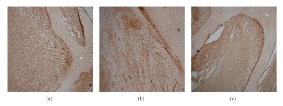
Immunohistochemistry for bone morphogenic protein (BMP; x100 magnification). (a) BMP-2, (b) BMP-4, and (c) BMP-7. The tumor cells stained positive for BMP-4 and negative for BMP-2 and BMP-7. *asterisk: bone tissue.
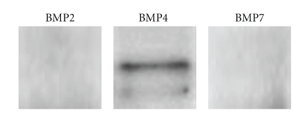
Western blot analysis for BMP. The tumor tissue expressed BMP-4 but not BMP-2 and -7.
Similar articles
-
Expression of bone morphogenic protein in sinonasal inverted papilloma with new bone formation.Allergy Rhinol (Providence). 2011 Jan;2(1):16-20. doi: 10.2500/ar.2011.2.0004. Allergy Rhinol (Providence). 2011. PMID: 22852110 Free PMC article.
-
Ancient schwannoma of the nasal septum associated with sphenoid sinus mucocele.Auris Nasus Larynx. 2010 Aug;37(4):522-5. doi: 10.1016/j.anl.2009.09.015. Epub 2010 Jan 4. Auris Nasus Larynx. 2010. PMID: 20045598
-
Sinonasal Schwannomas: Imaging Findings and Review of Literature.Ear Nose Throat J. 2023 Feb 12:1455613221150573. doi: 10.1177/01455613221150573. Online ahead of print. Ear Nose Throat J. 2023. PMID: 36775665 Review.
-
Nasal septal schwannoma: a rare sinonasal tumour with certain peculiarities.BMJ Case Rep. 2018 Apr 18;2018:bcr2017223850. doi: 10.1136/bcr-2017-223850. BMJ Case Rep. 2018. PMID: 29669769 Free PMC article. Review.
-
Sinonasal Schwannoma: A Case Report.Indian J Otolaryngol Head Neck Surg. 2024 Jun;76(3):2855-2858. doi: 10.1007/s12070-024-04547-5. Epub 2024 Feb 21. Indian J Otolaryngol Head Neck Surg. 2024. PMID: 38883519 Free PMC article.
Cited by
-
Ancient Schwannoma of the Thigh: Metaplastic Ossification, Cartilage Formation, and Extensive Calcification.Cureus. 2024 Feb 7;16(2):e53790. doi: 10.7759/cureus.53790. eCollection 2024 Feb. Cureus. 2024. PMID: 38465065 Free PMC article.
References
-
- Sheikh HY, Chakravarthy RP, Slevin NJ, Sykes AJ, Banerjee SS. Benign schwannoma in paranasal sinuses: a clinico-pathological study of five cases, emphasising diagnostic difficulties. Journal of Laryngology and Otology. 2008;122(6):598–602. - PubMed
-
- Yang TL, Hsu MC, Liu CM. Nasal schwannoma: a case report and clinicopathologic analysis. Rhinology. 2001;39(3):169–172. - PubMed
-
- Berlucchi M, Piazza C, Blanzuoli L, Battaglia G, Nicolai P. Schwannoma of the nasal septum: a case report with review of the literature. European Archives of Oto-Rhino-Laryngology. 2000;257(7):402–405. - PubMed
-
- Tosaka M, Hirato J, Miyagishima T, Saito N, Nakazato Y, Sasaki T. Calcified vestibular schwannoma with unusual histological characteristics—positive immunoreactivity for CD-34 antigen. Acta Neurochirurgica. 2002;144(4):395–399. - PubMed
-
- Atlas MD, Fagan PA, Turner J. Calcification of internal auditory canal tumors. Annals of Otology, Rhinology and Laryngology. 1992;101(7):620–622. - PubMed
Publication types
LinkOut - more resources
Full Text Sources
