. 2011:2011:785158.
doi: 10.1155/2011/785158. Epub 2010 Dec 9.

Mutation of herpesvirus Saimiri ORF51 glycoprotein specifically targets infectivity to hepatocellular carcinoma cell lines

Affiliations

Mutation of herpesvirus Saimiri ORF51 glycoprotein specifically targets infectivity to hepatocellular carcinoma cell lines

Susan J Turrell et al. J Biomed Biotechnol. 2011.

Abstract

Herpesvirus saimiri (HVS) is a gamma herpesvirus with several properties that make it an amenable gene therapy vector; namely its large packaging capacity, its ability to persist as a nonintegrated episome, and its ability to infect numerous human cell types. We used RecA-mediated recombination to develop an HVS vector with a mutated virion protein. The heparan sulphate-binding region of HVS ORF51 was substituted for a peptide sequence which interacts with somatostatin receptors (SSTRs), overexpressed on hepatocellular carcinoma (HCC) cells. HVS mORF51 showed reduced infectivity in non-HCC human cell lines compared to wild-type virus. Strikingly, HVS mORF51 retained its ability to infect HCC cell lines efficiently. However, neutralisation assays suggest that HVS mORF51 has no enhanced binding to SSTRs. Therefore, mutation of the ORF51 glycoprotein has specifically targeted HVS to HCC cell lines by reducing the infectivity of other cell types; however, the mechanism for this targeting is unknown.


Figures

Figure 1
Figure 1
Substitution of the heparan sulphate-binding region of HVS ORF51 protein. Analysis of the peptide sequence of the 269 amino acid protein indicates that there is an N-terminal signal sequence, 9 N-linked glycosylation sites, a potential heparan sulphate-binding site and a transmembrane domain. In the mutant ORF51 virus the heparan sulphate-binding site, highlighted in red, is replaced with the peptide sequence in blue. This sequence contains the SSTR binding motif.
Figure 2
Figure 2
Schematic of the RecA-mediated recombination method used to develop HVS mORF51. Two plasmids are transformed into competent E. coli cells that already harbour the HVS-GFP-BAC genome. These plasmids are both temperature sensitive and have antibiotic resistance markers for ease of selection. The first plasmid, pDF25-Tet, contains a RecA expression cassette to facilitate recombination, as well as a tetracycline resistance gene. The second, pKOV Kan, contains the mutated region of the HVS genome flanked on either side by regions of homology of at least 500 bp. These homology regions target the recombination event to a specific point in the viral DNA. pKOV Kan also contains a SacB gene, allowing negative selection on sucrose-containing medium, and a kanamycin resistance gene. When both plasmids are transformed into the E. coli, RecA expressed from pDF25-Tet induces a recombination event between one of the homology regions in pKOV Kan and the corresponding region in the HVS genome. Clones containing the pKOV Kan plasmid integrated into the HVS genome are then selected and made competent. These cointegrant clones are then retransformed with pDF25-Tet in order to produce a second recombination event. Depending on whether this recombination is in the same or adjacent homology region to the initial recombination, a revertant clone or a recombinant clone will be formed. Selection is used to identify recombinants, which can then be further analysed and confirmed by restriction digest and sequencing.
Figure 3
Figure 3
Possible orientations of pKOV Kan mORF51 cointegrants as seen by AgeI digest. (a) AgeI restriction map of the HVS genome. The restriction site marked in red is introduced when homologous recombination occurs between the pKOV Kan mORF51 plasmid and the complementary sequence in the viral genome. This introduced restriction site can then be used for identification of cointegrants. (b) Possible orientations of cointegrants. Depending on which homology region the reaction occurs (either 5′ or 3′ to the mutated ORF51 gene in pKOV Kan mORF51), cointegrants can be in one of two orientations. The mutated region containing the introduced AgeI site is shown in red. The pKOV Kan-mORF51 DNA is shown in blue, and the HVS DNA is shown in grey. The two orientations of cointegrant can be detected upon AgeI digestion analysis due to a 7 kb difference in the size of the ~50 kb fragment (see table). The larger ~80 kb fragment contains the terminal repeat (TR) region of the genome so is not suitable to detect this small difference as the number of TRs is not fixed.
Figure 4
Figure 4
AgeI restriction analysis of cointegrants formed by homologous recombination. The mutated version of the ORF51 gene contains an AgeI restriction site. This extra restriction site can be used as a marker for the integration of the pKOV Kan mORF51 vector into the HVS genome. Pulse field gel electrophoresis of possible cointegrant clones 3, 4, 8, and 9 reveals successful integration, as the ~130 kb band seen in HVS-GFP-BAC is cleaved into 2 smaller bands (indicated by arrows) due to the introduced restriction site. The 2 different orientations of pKOV Kan mORF51 can be visualised as clones 3 and 4 show a restriction pattern with bands at 45 kb and ~90 kb, whereas clones 8 and 9 have bands at 52 kb and ~85 kb. This 7 kb difference in the restriction site corresponds to the length of pKOV Kan mORF51.
Figure 5
Figure 5
Schematic describing the resolution of cointegrant clones. Cointegrants which have the pKOV Kan-mORF51 plasmid (shown in blue) inserted into the HVS genome (shown in grey) undergo a second recombination event, resulting in two possible outcomes. If the recombination site is in the same homology region as the first recombination, the intact pKOV Kan mORF51 plasmid is excised, forming a revertant. However, if this recombination occurs in the opposite homology region to the previous recombination event, the mutated region (shown in red) remains in the HVS genome, while the wild-type ORF51 is incorporated into the pKOV Kan plasmid.
Figure 6
Figure 6
AgeI restriction analysis of clones derived from cointegrants during homologous recombination. (a) Cointegrants formed from the first stage of RecA-mediated recombination underwent a second recombination event to remove the integrated pKOV Kan plasmid. This second recombination event could result in either a revertant or a mutant genotype. Of the 12 colonies screened, clone 9D had the desired restriction pattern consistent with a mutated ORF51 gene. (b) The mutant clone 9D was digested and run alongside a revertant 3A and the cointegrant clone 9 from which it originated. The loss of the integrated pKOV Kan plasmid can be visualised by the 52 kb band in the final lane decreasing in size by 7 kb to 45 kb.
Figure 7
Figure 7
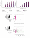
Comparison of wild-type and mutant virus entry measured by GFP expression. A range of human cancer cell lines were infected with increasing amounts of HVS-GFP-BAC (a), and HVS mORF51 (b). Owl monkey kidney (OMK) cells (permissive to the virus) were also used as a control, representative dot plots of which are shown in (c). 48 h after infection, GFP expression was measured by flow cytometry using a Becton Dickinson FacsCalibur (n = 2). Mutation of the ORF51 glycoprotein inhibits virus entry in OMK cells and several of the cancer cell lines. However, the HCC-derived cell lines are still able to be efficiently infected, suggesting that the SSTR binding region in the mutated protein facilitates viral attachement in these SSTR-expressing cells.
Figure 8
Figure 8
HVS mORF51 infection rates shown as a percentage of HVS-GFP-BAC infection in different cell types. Each virus, at an m.o.i of 2, was used to infect a panel of cell lines. GFP fluorescence (used as a marker of infected cells) was measured by flow cytometry 48 h after infection (n = 2). All hepatocellular carcinoma cell lines (Huh7, Huh7.5, and HepG2) have an infection rate of over 80% of that observed with HVS-GFP-BAC. In the permissive OMK cell line, as well as in the A549 lung cell line and HEK 293T cells, HVS mORF51 infects only approximately 20% of the number of cells infected with HVS-GFP-BAC. HVS mORF51 infects colorectal cancer cell line SW480 most inefficiently, producing an infection rate just of 4% compared to HVS-GFP-BAC.
Figure 9
Figure 9
Somatostatin receptor 1 is not required for HVS mORF51 cell entry. Two pancreatic cancer cell lines (MiaPaCa, and Panc1) were stably transfected with a construct expressing the SSTR1 gene. 48 h after infection with HVS mORF51, the cell lines expressing SSTR1 showed no increase in GFP expression as measured by flow cytometry, indicating that the recombinant virus does not interact with this receptor to enter the cell (n = 2).
Figure 10
Figure 10
Soluble heparin blocks HVS-GFP-BAC entry in Huh7.5 cells and inhibits HVS mORF51 to a lesser extent. Cells were incubated with a range of concentrations of heparin in 5% media for 1 h prior to infection with virus (also in 5% media). 48 h after infection, cells were removed from their wells, and GFP fluorescence was measured by flow cytometry (n = 2). Concentrations of heparin over 0.05 mg/ml resulted in viral cell entry being inhibited with both viruses. However, the neutralisation effect is less severe with HVS mORF51, suggesting some attenuation of its heparin-binding capacity.
Figure 11
Figure 11
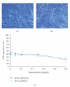
Somatostatin fails to neutralise HVS infection. The 14-amino acid peptide somatostatin was incubated with Huh7.5 cells at a range of concentrations for 1 h prior to infection with either HVS-GFP-BAC or HVS mORF51. After 48 h, GFP fluorescence was measured by flow cytometry (n = 2). Although cells incubated with up to 0.5 mg/ml somatostatin appeared healthy under the microscope (a), a concentration of 1 mg/ml was lethal (b). The graph (c) shows that both viruses exhibited negligible change in cell entry with increasing somatostatin concentration until the 0.5 mg/ml samples, where there is a reduced infection for both viruses. This may be due to the toxicity of the peptide rather than a true neutralisation effect.
Figure 12
Figure 12
SSTR antibody fails to neutralise HVS mORF51 infection. Huh7.5 cells were incubated with antibody (SSTR or mouse IgG negative control) for 1 h prior to infection with HVS-GFP-BAC or HVS mORF51. Samples were then measured for GFP expression using flow cytometry 48 h after infection (n = 2). Antibody concentration had no effect on either virus infection suggesting that SSTRs are not required for HVS mORF51 entry.

Similar articles

Cited by

References

    1. Roizman B, Carmichael LE, Deinhardt F. Herpesviridae. Definition, provisional nomenclature, and taxonomy. Intervirology. 1981;16(4):201–217. - PubMed
    1. Geevarghese SK, Geller DA, De Haan HA, et al. Phase I/II study of oncolytic herpes simplex virus NV1020 in patients with extensively pretreated refractory colorectal cancer metastatic to the liver. Human Gene Therapy. 2010;21(9):1119–1128. - PMC - PubMed
    1. Markert JM, Medlock MD, Rabkin SD, et al. Conditionally replicating herpes simplex virus mutant G207 for the treatment of malignant glioma: results of a phase I trial. Gene Therapy. 2000;7(10):867–874. - PubMed
    1. Senzer NN, Kaufman HL, Amatruda T, et al. Phase II clinical trial of a granulocyte-macrophage colony-stimulating factor-encoding, second-generation oncolytic herpesvirus in patients with unresectable metastatic melanoma. Journal of Clinical Oncology. 2009;27(34):5763–5771. - PubMed
    1. Macnab S, White R, Hiscox J, Whitehouse A. Production of an infectious Herpesvirus saimiri-based episomally maintained amplicon system. Journal of Biotechnology. 2008;134(3-4):287–296. - PubMed

Publication types

MeSH terms