Review
. 2011:2011:569034.
doi: 10.1155/2011/569034. Epub 2010 Dec 12.

Jun dimerization protein 2 controls senescence and differentiation via regulating histone modification

Affiliations
Review

Jun dimerization protein 2 controls senescence and differentiation via regulating histone modification

Yu-Chang Huang et al. J Biomed Biotechnol. 2011.

Abstract

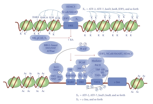
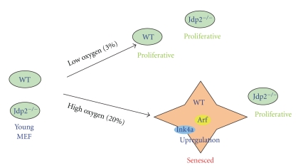
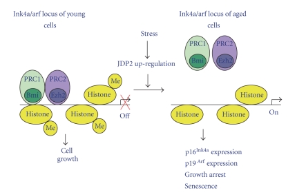
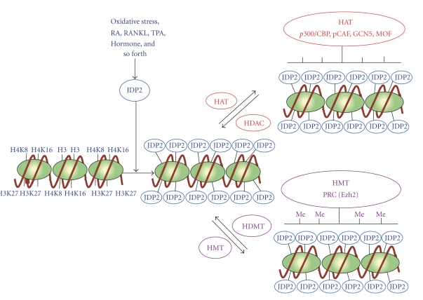
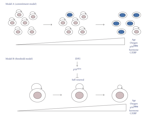
Transcription factor, Jun dimerization protein 2 (JDP2), binds directly to histones and DNAs and then inhibits the p300-mediated acetylation both of core histones and of reconstituted nucleosomes that contain JDP2 recognition DNA sequences. JDP2 plays a key role as a repressor of adipocyte differentiation by regulation of the expression of the gene C/EBPδ via inhibition of histone acetylation. Moreover, JDP2-deficient mouse embryonic fibroblasts (JDP2(-/-) MEFs) are resistant to replicative senescence. JDP2 inhibits the recruitment of polycomb repressive complexes (PRC1 and PRC2) to the promoter of the gene encoding p16(Ink4a), resulting from the inhibition of methylation of lysine 27 of histone H3 (H3K27). Therefore, it seems that chromatin-remodeling factors, including the PRC complex controlled by JDP2, may be important players in the senescence program. The novel mechanisms that underline the action of JDP2 in inducing cellular senescence and suppressing adipocyte differentiation are reviewed.


Figures

Figure 1
Figure 1
Schematic representation of the signal pathways of retinoic acid- (RA-) induced (RA-induced) differentiation of mouse embryonic carcinoma F9 cells. At the undifferentiation stage of F9 cells, HDAC3, NcoR/SMART, and JDP2 were recruited on the DRE (differentiation response element) in the promoter region of the c-jun gene to induce the heterochromatin. In the response to retinoic acid (RA), the signals of mitogen-activated kinase (MAPK; phosphorylated (p38(α/β)), BRG1-based SWI/SNF ATPase complex, Ini1/Snf5/SWI/SNF/BAF60 complex, p160 hormone coactivator (RARE/RXRE binding), and p300/PCAF complex were recruited to the DRE element of the c-Jun promoter and then Mediator, Pol II complex (IIB, IIF, IIE, IIH complex), TBP complex, and some TAF complex (ATF12, TAF 4 etc), and recruited and Pol II complex is elongated with phosphorylation of the carboxy terminal domain (CTD) of Pol II, and then c-Jun genes are finally activated. Ac, acetylated residues; K, lysine residues of histone; H3K, lysine residues of histone H3; H4K8, lysine residue at position 8 of histone H4; H4K16, lysine residue at position 16 of histone H4; X1 = ATF-2, ATF-7. JunD, JunB, JDP2, and so forth; X2 = c-Jun and so forth.
Figure 2
Figure 2
Oxydative stress controls the cell proliferation of mouse MEF cells through the epigenetic regulation of the p16Ink4a/Arf locus by JDP2. Young MEF primary cells exposed to oxidative stress (20%) accumulate JDP2. In the presence of JDP2, PRC1, and PRC2 dissociate from the p16Ink4a/Arf locus, and histone H3 on the promoter is demethylated. Finally, p16Ink4a and Arf are upregulated to express and then aged cells senescence. In the absence of JDP2, MEF cells do not enter the senescence stage and proliferate well. In the lower oxygen (3%), both WT and Jdp2−/− MEF cells are not senesced but proliferated.
Figure 3
Figure 3
Proposed model of the epigenetic regulation of the expression of the genes for p16Ink4a and Arf by JDP2. The exposure of young MEF primary cells to aging stress leads to the accumulation of JDP2. JDP2 binds to histones and inhibits the methylation of H3K27 at the p16Ink4a/Arf locus. As a result, PRC-1 and PRC-2 fail to form stable repressive complexes and are released from the locus. The consequent expression of p16Ink4a and Arf in the aged cells leads to growth arrest and senescence stage.
Figure 4
Figure 4
Model for the epigenetic regulation by JDP2. During the exposure of cells with oxydative stress, retinoic acid (RA), RANKL, TPA, and adipocyte inducing hormones, the histone H3, H4K8, and H4K16 as well as H3K27 were masked by JDP2 proteins and prevent the attack of histone modification enzymes like HAT (p300/CBP, pCAF, GCN5, MOF, etc.) and HMT (Ezh2 etc) in the cellular senescence and cell differentiation. This is a novel mechanism of JDP2 to inhibit the histone modification. The gene locus is either the p16Ink4a/Arf locus or C/EBPδ locus.
Figure 5
Figure 5
Model of role of JDP2 in cellular aging and cell differentiation. Self-renewing cells undergo repeated divisions, and p16Ink4aor C/EBPδ expression increases with age, oxygen, and hormone, as a consequence of undefined stimuli. (a) Commitment model: p16Ink4a or C/EBPδ expression occurs stochastically in a subpopulation of cells resulting in their senescence. In this model, the capacity for self-renewal of the noncommitted cells is not affected. (b) Threshold model: expression of p16Ink4a or C/EBPδ increases or decreases, respectively, uniformly in the tissue specific. Self-renewing cells compartment is compromised over time. Self-renewal is indicated by curved arrows; committed cells are colored blue.

References

    1. Kouzarides T. Chromatin modifications and their function. Cell. 2007;128(4):693–705. - PubMed
    1. Shilatifard A. Chromatin modifications by methylation and ubiquitination: implications in the regulation of gene expression. Annual Review of Biochemistry. 2006;75:243–269. - PubMed
    1. Li B, Carey M, Workman JL. The role of chromatin during transcription. Cell. 2007;128(4):707–719. - PubMed
    1. Ekwall K. Genome-wide analysis of HDAC function. Trends in Genetics. 2005;21(11):608–615. - PubMed
    1. Luger K, Mäder AW, Richmond RK, Sargent DF, Richmond TJ. Crystal structure of the nucleosome core particle at 2.8 Å resolution. Nature. 1997;389(6648):251–260. - PubMed

Publication types

LinkOut - more resources