Review
. 2011:36:31-59.
doi: 10.1007/978-3-7091-0179-7_3.

Gustatory and reward brain circuits in the control of food intake

Affiliations
Review

Gustatory and reward brain circuits in the control of food intake

A J Oliveira-Maia et al. Adv Tech Stand Neurosurg. 2011.

Abstract

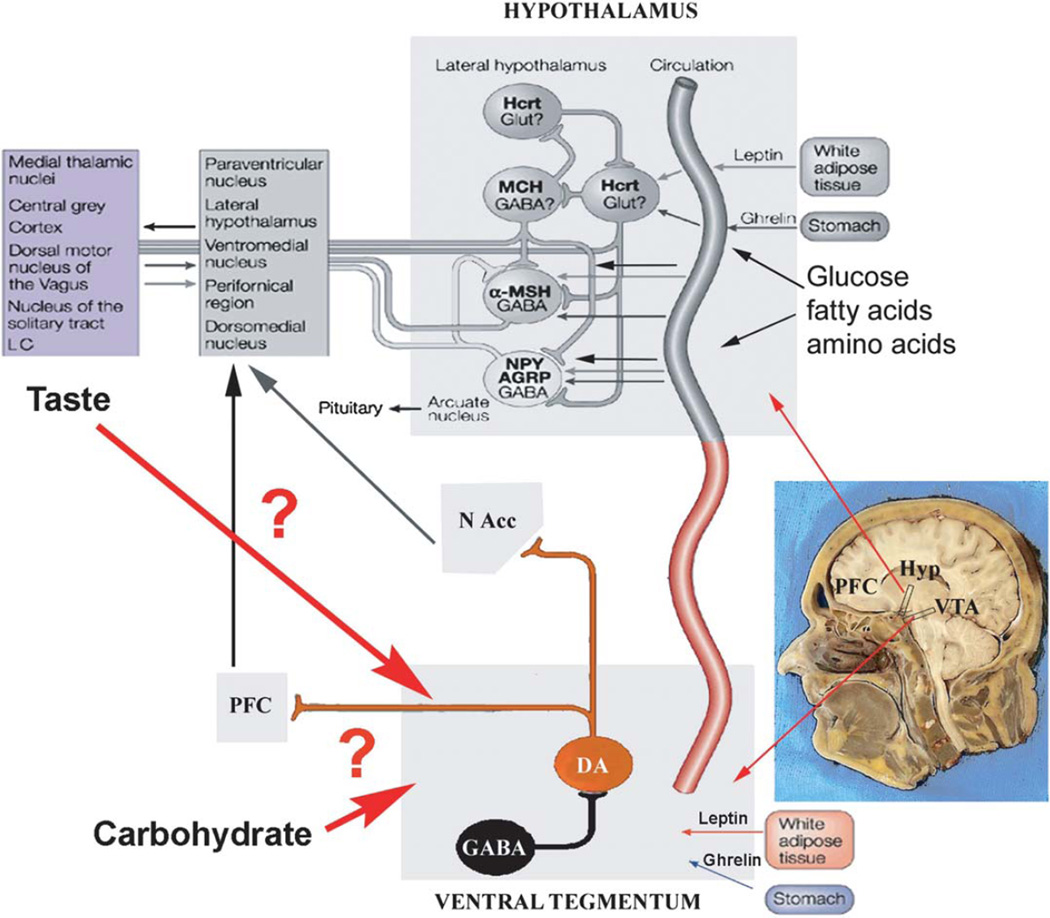
Gustation is a multisensory process allowing for the selection of nutrients and the rejection of irritating and/or toxic compounds. Since obesity is a highly prevalent condition that is critically dependent on food intake and energy expenditure, a deeper understanding of gustatory processing is an important objective in biomedical research. Recent findings have provided evidence that central gustatory processes are distributed across several cortical and subcortical brain areas. Furthermore, these gustatory sensory circuits are closely related to the circuits that process reward. Here, we present an overview of the activation and connectivity between central gustatory and reward areas. Moreover, and given the limitations in number and effectiveness of treatments currently available for overweight patients, we discuss the possibility of modulating neuronal activity in these circuits as an alternative in the treatment of obesity.


Figures

Fig. 1
Fig. 1. Peripheral Taste Mechanisms
Tastants activate two classes of taste bud cells: Type II or receptor cells and Type III or presynaptic cells. Different subclasses of receptor cells (green, red, and blue cells), express T1R2/T1R3, T2R or T1R1/T1R3 G-protein-coupled taste receptors and are activated respectively by sweet, bitter or umami compounds. Downstream signaling pathways in these cells require phospholipase C β2 and transient receptor potential ion channel M5 (TRPM5). When activated, receptor cells release adenosine triphosphate (ATP), which is then thought to act upon intragemmal taste nerve fibers (black fibres) and/or presynaptic cells. Presypnaptic cells (purple cell) express synapse-related proteins such as synaptosomal-associated protein of 25kD and form conventional synapses with intragemmal processes of peripheral taste neurons. In contrast with receptor cells, presynaptic cells are broadly tuned to tastants of multiple qualities – currently, they are thought to be activated directly by sour stimuli, through a different set of receptors and signaling pathways than those used by receptor cells, and indirectly by sweet, bitter and umami compounds, through ATP released from receptor cells. Serotonin (5-HT) is also released from taste buds upon chemosensory stimulation, presumably in synapses between receptor cells and taste neurons. Several taste bud cell types, including receptor cells and type I cells, have been proposed to transduce salt stimuli, but there is still no consensus (see text; adapted from Tomchik, Berg et al. 2007, used with permission).
Fig. 2
Fig. 2. Gut Nutrient Signaling Pathways
Ingested nutrients elicit mechanosensory and chemosensory responses in the gut, as represented in green on the right. Postingestive responses depend mainly on the production of gut hormones, such as CCK and GLP-1, that signal nutrient presence and quality by activating vagal afferents (blue, dashed lines) and/or entering blood circulation via the portal vein (red, solid lines). Absorbed nutrients (glucose and other ‘fuels’) and feeding-related peptides produced in sites other than the gut (liver, muscle, adipose tissue and pancreas, on the bottom left), are two other categories of gustatory humoral signals. The postingestive sensory information thus generated, modulates the activity of central neural circuits at several levels of the brain, represented on the top left (from Zheng and Berthoud 2008, used with permission).
Fig. 3
Fig. 3. Anatomy of the main central gustatory pathways
Taste-specific information is conveyed by cranial nerves VII, IX and X (blue lines) to the rostral division of the solitary tract nucleus (rNTS) in the medulla. In primates, fibres (red lines) from second-order taste neurons in the rNTS project ipsilaterally to the parvicellular division of the ventral posterior nucleus of the thalamus (VPpc). Thalamic efferents (green lines) then project to the insula, defining the primary gustatory cortex which, in turn, projects (black lines) to the orbitofrontal cortex, sometimes defined as a secondary cortical taste area. The parabrachial nuclei (PbN) of the pons are shown in orange. In rodents these are a relay for taste afferents from the rNTS. In both primates and rodents, the PbN also receive second order visceral sensory fibres from the caudal division of the solitary tract nucleus (cNTS), transmitted mainly through the vagus nerve (not shown). The PbN has a dorsal thalamocortical projection to the VPMpc and also a ventral projection that terminates in amygdalar and hypothalamic nuclei, among others (adapted from Simon, de Araujo et al. 2006, used with permission).
Fig. 4
Fig. 4. Hedonic and homeostatic regulation of feeding
Current literature considers the hypothalamus as the main centre for feeding regulation. Lateral hypothalamus neurons that produce orexin (also known as hypocretin - Hcrt) and melanin concentrating hormone (MCH) are potent stimulators of food intake. Neurons in the arcuate nucleus of the hypothalamus synthesize melanocyte stimulating hormone (MSH) or neuropeptide Y (NPY) that have opposed effects in the control of food intake and energy expenditure. The hypothalamic nuclei are traditionally considered homeostatic centre for feeding regulation since they respond to peripheral metabolic hormones and fuels (such as leptin and ghrelin) that are critical for energy homeostasis (Gao and Horvath 2007). The mesencephalic dopamine system, on the other hand, responds robustly to a diverse array of rewarding stimuli, including food, and plays a critical role in the behavioural responses to these stimuli (Wise 2006). Orosensory responses to palatable food are sufficient for the occurrence of dopamine (DA) responses in the mesolimbic system (Hajnal, Smith et al. 2004), which have generally been considered as a system for ‘hedonic’ regulation of food intake. However, some of the peripheral hormones that modulate the behavioural components of energy homeostasis also impact the activity in this system (see text). Furthermore, in a recent publication, de Araujo and Oliveira-Maia et al (de Araujo, Oliveira-Maia et al. 2008) have shown, using ‘taste-blind’ mice (Zhang, Hoon et al. 2003), that the caloric value of sucrose, in the absence of taste transduction, is also sufficient to activate the midbrain reward circuitry. While the physiological details of the signaling mechanisms involved remain to be described, it seems reasonable to suggest that the distinction between hedonic and homeostatic regulation of feeding is redundant. GABA, gamma-aminobutyric acid; Glut, glutamate; Hyp, hypothalamus, NAcc, nucleus accumbens; PFC, prefrontal cortex, VTA, ventral tegmental area (from Andrews and Horvath 2008, used with permission from Elsevier).

References

    1. Abbott CR, Monteiro M, et al. The inhibitory effects of peripheral administration of peptide YY(3-36) and glucagon-like peptide-1 on food intake are attenuated by ablation of the vagal-brainstem-hypothalamic pathway. Brain Res. 2005;1044(1):127–131. - PubMed
    1. Abizaid A, Liu ZW, et al. Ghrelin modulates the activity and synaptic input organization of midbrain dopamine neurons while promoting appetite. J Clin Invest. 2006;116(12):3229–3339. - PMC - PubMed
    1. Accolla R, Bathellier B, et al. Differential spatial representation of taste modalities in the rat gustatory cortex. J Neurosci. 2007;27(6):1396–1404. - PMC - PubMed
    1. Altschuler SM, Bao XM, et al. Viscerotopic representation of the upper alimentary tract in the rat: sensory ganglia and nuclei of the solitary and spinal trigeminal tracts. J Comp Neurol. 1989;283(2):248–268. - PubMed
    1. Anand BK, Brobeck JR. Localization of a "feeding center" in the hypothalamus of the rat. Proc Soc Exp Biol Med. 1951;77(2):323–324. - PubMed

Publication types

LinkOut - more resources