Review
. 2011 Sep-Oct;3(5):606-18.
doi: 10.1002/wsbm.133. Epub 2010 Dec 31.

Ischemic kidney injury and mechanisms of tissue repair

Affiliations
Review

Ischemic kidney injury and mechanisms of tissue repair

Marwa El Sabbahy et al. Wiley Interdiscip Rev Syst Biol Med. 2011 Sep-Oct.

Abstract

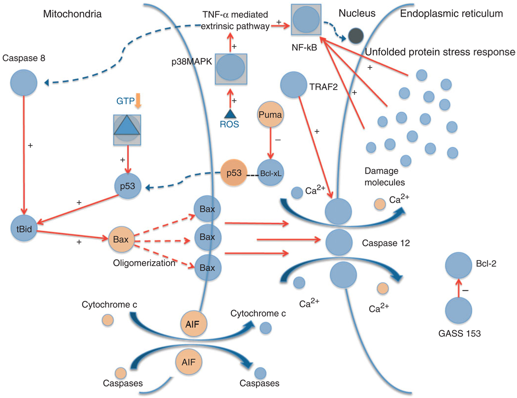
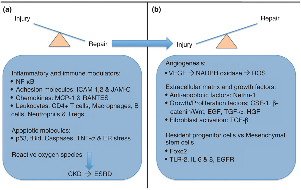
Acute kidney injury (AKI) may result from ischemia or by the use of nephrotoxic agents. The incidence of AKI is variable, depends on comorbidities, and ranges from 5 to 35% in all hospitalized patients. The mechanisms of kidney injury exist within a large network of signaling pathways driven by interplay of inflammatory cytokines/chemokines, reactive oxygen species (ROS), and apoptotic factors. The effects and progression of injury overlap extensively with the remarkable ability of the kidney to repair itself both by intrinsic and extrinsic mechanisms that involve specific cell receptors/ligands as well as possible paracrine influences. The fact that kidney injury is usually part of a generalized comorbid condition makes it all the more challenging in terms of assessment of severity. In this review, we attempt to analyze the mechanisms of ischemic injury and repair in acute and chronic kidney disease from the perspectives of both preclinical and human studies.


Figures

FIGURE 1
FIGURE 1
Kidney tissue injury (a–d) and repair (e–h) over time following 20 min of bilateral renal ischemia/reperfusion injury. Male Wistar rats were subjected to sham or bilateral ischemia by clamping the renal pedicles for 20 min and then removing the clamps and confirming reperfusion. Rats were euthanized at various times and kidney tissues were collected. Representative photomicrographs of H&E-stained paraffin-embedded kidney sections (at 200 × magnification) and immunohistochemistry for Ki67 (at 400 × magnification) are presented from the following time points: (a, e) Sham surgery; (b, f) 24 h; (c, g) 72 h; and (d, h) 120 h. All fields were chosen from the cortex and outer medulla. Arrows in panels b and c indicate sloughing of cells, tubular dilation and necrosis. Arrows in panels e–h show Ki67 positive nuclei as an indicator of tubular epithelial cell proliferation.
FIGURE 2
FIGURE 2
Apoptosis in the kidney tissue over time following 20 min of bilateral renal ischemia/reperfusion injury. Male Wistar rats were subjected to sham or bilateral ischemia by clamping the renal pedicles for 20 min and then removing the clamps and confirming reperfusion. Rats were euthanized at various times, kidney tissues were collected and transferase dUTP nick end labeling (TUNEL) immunostaining was performed to label apoptotic cells. Representative photomicrographs at 400 × magnification are presented. Arrows in panels show TUNEL positive nuclei as an indicator of tubular epithelial cell apoptosis.
FIGURE 3
FIGURE 3
Apoptosis: intrinsic, extrinsic, and endoplasmic reticulum (ER) stress mechanisms. The diagram shows the various signaling pathways leading to apoptosis and the generation of ROS. Upon stress activation, p53 translocates to the mitochondria where it interacts with the BcL-2 family members, including anti-apoptotic BcL-xL and pro-apoptotic Bax and Bak. P53 disrupts inhibitory complexes formed with tBid and Bim, which effectively activate Bax and Bak to oligomerize and form lipid pores in the outer mitochondrial membrane, a process called mitochondrial outer membrane permeabilization (MOMP). The Bax/Bak pathway subsequently activates caspase 12 mediated apoptosis through the release of Ca2+ into the cytoplasm. Cytoplasmic p53 also plays a role in transcription independent apoptosis by being liberated from BcL-xL by Puma, and then activating cytosolic Bax. TNF-α is responsible for extrinsic death receptor mediated apoptosis, which is proposed to be activated by ROS stimulation through the p38 MAPK pathway. This leads to NF-κB and caspase 8 activation, which link the extrinsic pathway to the intrinsic apoptotic mechanism initiated by p53. The ER stress response mechanism might be triggered by the accumulation of ‘damage molecules’ in the ER, stimulating the unfolded protein response (UPR) and activating NF-κB. GADD-153 activates pro-apoptotic factors and downregulates anti-apoptotic BcL-2. Tumor necrosis factor receptor-associated factor-2 (TRAF-2) and caspase 12 stimulation contribute to apoptosis and cell death.
FIGURE 4
FIGURE 4
Overview of injury and repair mechanisms. In the early stages of kidney injury, there is involvement of numerous inflammatory and immune modulators, including NF-κB, adhesion molecules such as ICAM and junctional adhesion molecule (JAM)-C, chemokines, neutrophils, and CD4+ T cells. Apoptotic molecules including p53, caspases, and tBid play a corresponding role along with these inflammatory factors. The late stages of injury involve ‘clean house’ molecules including mainly Tregs and macrophages in preparation for tissue recovery. If the insult is severe and/or prolonged it may progress to chronic kidney disease (CKD), driven by the production of free radicals and activated fibroblasts. Reparative mechanisms include angiogenic factors such as vascular endothelial growth factor (VEGF), anti-apoptotic factors like Netrin-1, and growth/proliferation factors such as Wingless (Wnt)/β-catenin, and transforming growth factor (TGF)-α and -β. Transcription factors including the Fox family, as well as Toll-like receptors (TLR)-2 and interleukin (IL)-6, -8 are involved in the influences of adult/renal progenitor stem cells and mesenchymal cells on repair.

References

    1. Kellum JA. Acute kidney injury. Crit Care Med. 2008;36(4 suppl):S141–S145. - PubMed
    1. Soni SS, Ronco C, Katz N, Cruz DN. Early diagnosis of acute kidney injury: the promise of novel biomarkers. Blood Purif. 2009;28:165–174. - PubMed
    1. Haase M, Bellomo R, Matalanis G, Calzavacca P, Dragun D, Haase-Fielitz A. A comparison of the RIFLE and Acute Kidney Injury Network classifications for cardiac surgery-associated acute kidney injury: a prospective cohort study. J Thorac Cardiovasc Surg. 2009;138:1370–1376. - PubMed
    1. Murray PT, Devarajan P, Levey AS, Eckardt KU, Bonventre JV, Lombardi R, Herget-Rosenthal S, Levin A. A framework and key research questions in AKI diagnosis and staging in different environments. Clin J Am Soc Nephrol. 2008;3:864–868. - PubMed
    1. Waikar SS, Bonventre JV. Creatinine kinetics and the definition of acute kidney injury. J Am Soc Nephrol. 2009;20:672–679. - PMC - PubMed

FURTHER READING

    1. Brenner BM, Rector FC. Brenner & Rector’s the Kidney. 8th ed. Philadelphia, PA: Saunders Elsevier; 2008.
    1. Schrier RW. Manual of Nephrology. 7th ed. Philadelphia, PA: Lippincott Williams & Wilkins; 2009.

Publication types