. 2011 May;163(2):283-98.
doi: 10.1111/j.1476-5381.2010.01187.x.

γ-Tocotrienol is a novel inhibitor of constitutive and inducible STAT3 signalling pathway in human hepatocellular carcinoma: potential role as an antiproliferative, pro-apoptotic and chemosensitizing agent

Affiliations

γ-Tocotrienol is a novel inhibitor of constitutive and inducible STAT3 signalling pathway in human hepatocellular carcinoma: potential role as an antiproliferative, pro-apoptotic and chemosensitizing agent

Peramaiyan Rajendran et al. Br J Pharmacol. 2011 May.

Abstract

Background and purpose: Activation of signal transducer and activator of transcription 3 (STAT3) play a critical role in the survival, proliferation, angiogenesis and chemoresistance of tumour cells. Thus, agents that suppress STAT3 phosphorylation have potential as cancer therapies. In the present study, we investigated whether the apoptotic, antiproliferative and chemosensitizing effects of γ-tocotrienol are associated with its ability to suppress STAT3 activation in hepatocellular carcinoma (HCC).

Experimental approach: The effect of γ-tocotrienol on STAT3 activation, associated protein kinases and phosphatase, STAT3-regulated gene products, cellular proliferation and apoptosis in HCC cells was investigated.

Key results: γ-Tocotrienol inhibited both the constitutive and inducible activation of STAT3 with minimum effect on STAT5. γ-Tocotrienol also inhibited the activation of Src, JAK1 and JAK2 implicated in STAT3 activation. Pervanadate reversed the γ-tocotrienol-induced down-regulation of STAT3, suggesting the involvement of a protein tyrosine phosphatase. Indeed, we found that γ-tocotrienol induced the expression of the tyrosine phosphatase SHP-1 and deletion of the SHP-1 gene by small interfering RNA abolished the ability of γ-tocotrienol to inhibit STAT3 activation. γ-Tocotrienol also down-regulated the expression of STAT3-regulated gene products, including cyclin D1, Bcl-2, Bcl-xL, survivin, Mcl-1 and vascular endothelial growth factor. Finally, γ-tocotrienol inhibited proliferation, induced apoptosis and significantly potentiated the apoptotic effects of chemotherapeutic drugs (paclitaxel and doxorubicin) used for the treatment of HCC.

Conclusions and implications: Overall, these results suggest that γ-tocotrienol is a novel blocker of the STAT3 activation pathway, with a potential role in future therapies for HCC and other cancers.


Figures

Figure 1
Figure 1
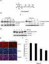
γ-Tocotrienol inhibits constitutively active signal transducer and activator of transcription 3 (STAT3) in HepG2 cells. (A) The structure of γ-tocotrienol. (B) γ-Tocotrienol suppresses phospho-STAT3 levels in a dose-dependent manner. HepG2 cells (2 × 10⁶ mL−1) were treated with the indicated concentrations of γ-tocotrienol for 4 h, after which whole-cell extracts were prepared, and 30 µg of protein was resolved on 7.5% SDS-PAGE gel, electrotransferred onto nitrocellulose membranes, and probed for phospho-STAT3. (C) γ-Tocotrienol suppresses phospho-STAT3 levels in a time-dependent manner. HepG2 cells were treated with the 50 µM γ-tocotrienol for the indicated times, after which Western blotting was performed as described for (B). (D) HepG2 cells were treated with 50 µM γ-tocotrienol for the indicated times. Whole-cell extracts were prepared, fractionated on SDS-PAGE, and examined by Western blotting. (E) γ-Tocotrienol causes inhibition of translocation of STAT3 to the nucleus. HepG2 cells (1 × 10⁵ mL−1) were incubated with or without 50 µM γ-tocotrienol for 6 h and then analysed for the intracelullar distribution of STAT3 by immunocytochemistry. The same slides were counterstained for nuclei with Hoechst (50 ng·mL−1) for 5 min. Each panel is representative of three independent experiments. (F) γ-Tocotrienol suppresses NF-κB activation in HepG2 cells. HepG2 cells were treated with indicated concentrations of γ-tocotrienol for 6 h; nuclear extracts were prepared, and 20 µg of the nuclear extract protein was used for elisa-based DNA binding assay as described in Methods. The results shown are representative of three independent experiments. **P < 0.01, ***P < 0.001.
Figure 2
Figure 2
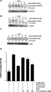
γ-Tocotrienol down-regulates IL-6-induced phospho-STAT3 (signal transducer and activator of transcription 3) activation in hepatocellular carcinoma cells. (A) SNU-387 (2 × 10⁶ mL−1) were treated with 50 µM γ-tocotrienol for the indicated times and then stimulated with IL-6 (10 ng·mL−1) for 15 min. Whole-cell extracts were then prepared and analysed for phospho-STAT3 by Western blotting. The same blots were stripped and reprobed with STAT3 antibody to verify equal protein loading. The results shown are representative of three independent experiments. (B) SNU-387 (2 × 10⁶ mL−1) were treated with 50 µM γ-tocotrienol for the indicated times and then stimulated with IL-6 (10 ng·mL−1) for 15 min. Whole-cell extracts were then prepared and analysed for phospho-JAK2 by Western blotting. The same blots were stripped and reprobed with JAK2 antibody to verify equal protein loading. (C) SNU-387 (2 × 10⁶ mL−1) were treated with 50 µM γ-tocotrienol for the indicated times and then stimulated with IL-6 (10 ng·mL−1) for 15 min. Whole-cell extracts were then prepared and analysed for phospho-Akt by Western blotting. The same blots were stripped and reprobed with Akt antibody to verify equal protein loading. (D) PLC/PRF-5 cells (5 × 10⁵ mL−1) were transfected with STAT3-luciferase (STAT3-Luc) plasmid, incubated for 24 h, and treated with 10, 25 and 50 µM γ-tocotrienol for 6 h and then stimulated with epidermal growth factor (EGF) (100 ng·mL−1) for 2 h. Whole-cell extracts were then prepared and analysed for luciferase activity. The results shown are representative of three independent experiments. * Indicates P < 0.05, comparison between EGF- and γ-tocotrienol-treated groups by Student's t-test.
Figure 3
Figure 3
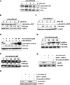
(A) γ-Tocotrienol suppresses phospho-Src levels. HepG2 cells (2 × 10⁶ mL−1) were treated with 50 µM γ-tocotrienol, after which whole-cell extracts were prepared and Western blotting was performed. (B) γ-Tocotrienol suppresses phospho-JAK1 levels. HepG2 cells were treated with 50 µM γ-tocotrienol for indicated time intervals, after which whole-cell extracts were prepared and Western blotting was performed. (C) γ-Tocotrienol suppresses phospho-JAK2 levels. HepG2 cells were treated with 50 µM γ-tocotrienol for indicated time intervals, after which whole-cell extracts and Western blotting was performed. (D) Pervanadate reverses the phospho-STAT3 (signal transducer and activator of transcription 3) inhibitory effect of γ-tocotrienol. HepG2 cells were treated with the indicated concentrations of pervanadate and 50 µM γ-tocotrienol for 6 h, after which Western blotting was performed. (E) γ-Tocotrienol induces the expression of SHP-1 protein in HepG2 cells. HepG2 cells were treated with indicated concentrations of γ-tocotrienol for 6 h, after which Western blotting was performed. (F) Effect of γ-tocotrienol on SHP-1 mRNA expression in HepG2 cells. HepG2 cells were treated with indicated concentrations of γ-tocotrienol for 6 h, after which RNA samples were subjected to RT-PCR with SHP-1 and GAPDH specific primers. PCR products were run on 1% agarose gel containing GelRed. Stained bands were visualized under UV light and photographed. (G) Effect of SHP-1 knockdown on γ-tocotrienol induced expression of SHP-1. HepG2 cells were transfected with either SHP-1 siRNA or scrambled siRNA (50 nM). After 24 h, cells were treated with 50 µM γ-tocotrienol for 6 h and whole-cell extracts were subjected to Western blot analysis. (H) HepG2 cells were transfected with either SHP-1 siRNA or scrambled siRNA (50 nM). After 24 h, cells were treated with 50 µM γ-tocotrienol for 6 h and whole-cell extracts were subjected to Western blot analysis for phosphorylated STAT3. The results shown are representative of three independent experiments.
Figure 4
Figure 4
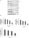
(A) γ-Tocotrienol suppresses signal transducer and activator of transcription 3 (STAT3) regulated gene products involved in proliferation, survival and angiogenesis. HepG2 cells (2 × 10⁶ mL−1) were treated with 25 µM γ-tocotrienol for indicated time intervals, after which whole-cell extracts were prepared and 30 µg portions of those extracts were resolved on 10% SDS-PAGE, membrane sliced according to molecular weight and probed against cyclin D1, Bcl-2, Bcl-xL, survivin, Mcl-1 and vascular endothelial growth factor (VEGF) antibodies. The same blots were stripped and reprobed with β-actin antibody to verify equal protein loading. The results shown are representative of three independent experiments. (B) HepG2 cells (3 × 10⁵ mL−1) were treated with 25 µM γ-tocotrienol for the indicated time intervals, after which cells were harvested after treatment and RNA samples, were extracted; 1 µg portions of the respective RNA extracts then proceed for reverse transcription to generate corresponding cDNA. Real-time PCR was performed to measure the relative quantities of mRNA. cDNA product was targeted against cyclin D1, Bcl-xL and Mcl-1 TaqMan probes, with HuGAPDH as endogenous control for measurement of equal loading of RNA samples. Results were analysed using Sequence Detection Software version 1.3 provided by Applied Biosystems. Gene expression was normalized with endogenous HuGAPDH by determination of the difference in threshold cycle (Ct) between treated and untreated cells using 2-ΔΔCt method. Relative gene expression was expressed as fold change of treated samples against untreated. Untreated samples were set at a value of one. Values represent mean ± SEM. *P < 0.05, indicates comparison between untreated and γ-tocotrienol-treated groups for 4, 6, 8 and 12 h by Student's t-test.
Figure 5
Figure 5
γ-Tocotrienol suppresses proliferation and activates caspase-3. (A) HepG2, C3A and SNU-387 cells (5 × 10³ mL−1) were plated in triplicate, treated with indicated concentrations of γ-tocotrienol, and then subjected to 3-(4, 5-dimethylthiazol-2-yl)-2, 5-diphenyl-2H-tetrazolium bromide assay after 24, 48 and 72 h to analyse proliferation of cells. Vertical lines indicate SD between the triplicates. *P < 0.05, comparison between untreated and γ-tocotrienol-treated groups for 24, 48 and 72 h by Student's t-test. (B) HepG2 cells were treated with 25 µM γ-tocotrienol for the indicated times, whole-cell extracts were prepared, separated on SDS-PAGE, and subjected to Western blotting against procaspase-3 antibody. The same blot was stripped and reprobed with β-actin antibody to show equal protein loading. (C) HepG2 cells were treated with 25 µM γ-tocotrienol for the indicated times, and whole-cell extracts were prepared, separated on SDS-PAGE, and subjected to Western blot against PARP antibody. The same blot was stripped and reprobed with β-actin antibody to show equal protein loading. Densitiometric analysis for 116 kDa band of PARP was performed using Image J software. The results shown are representative of three independent experiments.
Figure 6
Figure 6
(A) Overexpression of constitutive signal transducer and activator of transcription 3 (STAT3) protects Hep3B cells from γ-tocotrienol-induced cytotoxicity. Hep3B cells were transfected with constitutive STAT3 plasmid. After 24 h of transfection, the cells were treated with 50 µM γ-tocotrienol for 24 h, and then the cytotoxicity was determined by the live/dead assay and 20 random fields were counted. (B) γ-Tocotrienol potentiates the apoptotic effect of doxorubicin and paclitaxel. HepG2 cells (1 × 10⁶ mL−1) were treated with 10 µM γ-tocotrienol and 10 nM doxorubicin or 5 nM paclitaxel alone or in combination for 48 h at 37°C. Cells were stained with a live/dead assay reagent for 30 min and then analysed under a fluorescence microscope as described in Methods. The results shown are % apoptosis and are representative of three independent experiments.

Similar articles

Cited by

References

    1. Aggarwal BB, Sethi G, Ahn KS, Sandur SK, Pandey MK, Kunnumakkara AB, et al. Targeting signal-transducer-and-activator-of-transcription-3 for prevention and therapy of cancer: modern target but ancient solution. Ann N Y Acad Sci. 2006;1091:151–169. - PubMed
    1. Aggarwal BB, Kunnumakkara AB, Harikumar KB, Gupta SR, Tharakan ST, Koca C, et al. Signal transducer and activator of transcription-3, inflammation, and cancer: how intimate is the relationship? Ann N Y Acad Sci. 2009a;1171:59–76. - PMC - PubMed
    1. Aggarwal BB, Vijayalekshmi RV, Sung B. Targeting inflammatory pathways for prevention and therapy of cancer: short-term friend, long-term foe. Clin Cancer Res. 2009b;15:425–430. - PubMed
    1. Aggarwal BB, Sundaram C, Prasad S, Kannapan R. Tocotrienols, the vitamin E of the 21st century: its potential against cancer and other chronic diseases. Biochem Pharmacol. 2010;80:1613–1631. - PMC - PubMed
    1. Ahn KS, Sethi G, Krishnan K, Aggarwal BB. Gamma-tocotrienol inhibits nuclear factor-kappaB signaling pathway through inhibition of receptor-interacting protein and TAK1 leading to suppression of antiapoptotic gene products and potentiation of apoptosis. J Biol Chem. 2007;282:809–820. - PubMed

Publication types

MeSH terms