. 2011 Nov;15(11):2430-42.
doi: 10.1111/j.1582-4934.2010.01246.x.

Caveolin-1 is required for contractile phenotype expression by airway smooth muscle cells

Affiliations

Caveolin-1 is required for contractile phenotype expression by airway smooth muscle cells

Reinoud Gosens et al. J Cell Mol Med. 2011 Nov.

Abstract

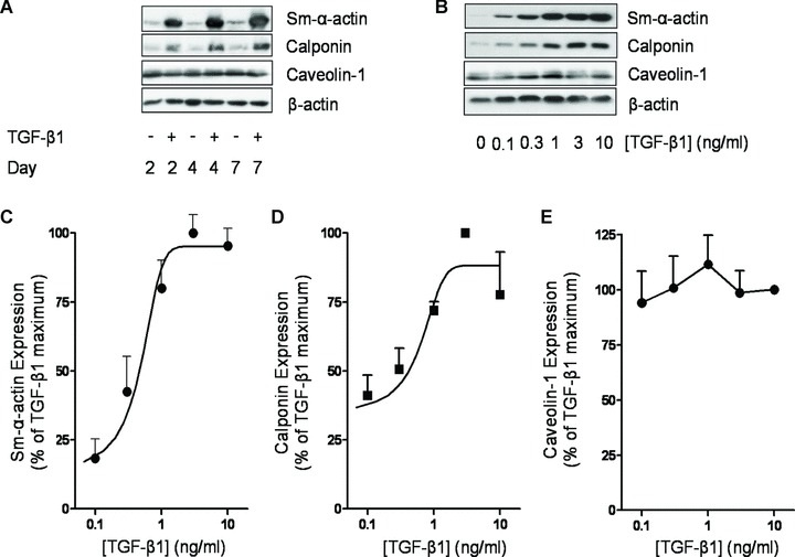
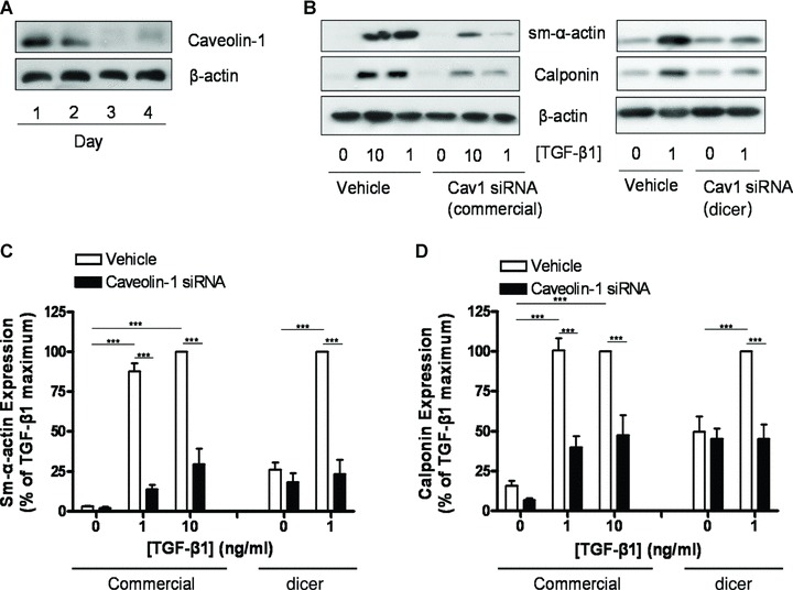
Airway smooth muscle cells exhibit phenotype plasticity that underpins their ability to contribute both to acute bronchospasm and to the features of airway remodelling in chronic asthma. A feature of mature, contractile smooth muscle cells is the presence of abundant caveolae, plasma membrane invaginations that develop from the association of lipid rafts with caveolin-1, but the functional role of caveolae and caveolin-1 in smooth muscle phenotype plasticity is unknown. Here, we report a key role for caveolin-1 in promoting phenotype maturation of differentiated airway smooth muscle induced by transforming growth factor (TGF)-β(1). As assessed by Western analysis and laser scanning cytometry, caveolin-1 protein expression was selectively enriched in contractile phenotype airway myocytes. Treatment with TGF-β(1) induced profound increases in the contractile phenotype markers sm-α-actin and calponin in cells that also accumulated abundant caveolin-1; however, siRNA or shRNAi inhibition of caveolin-1 expression largely prevented the induction of these contractile phenotype marker proteins by TGF-β(1). The failure by TGF-β(1) to adequately induce the expression of these smooth muscle specific proteins was accompanied by a strongly impaired induction of eukaryotic initiation factor-4E binding protein(4E-BP)1 phosphorylation with caveolin-1 knockdown, indicating that caveolin-1 expression promotes TGF-β(1) signalling associated with myocyte maturation and hypertrophy. Furthermore, we observed increased expression of caveolin-1 within the airway smooth muscle bundle of guinea pigs repeatedly challenged with allergen, which was associated with increased contractile protein expression, thus providing in vivo evidence linking caveolin-1 expression with accumulation of contractile phenotype myocytes. Collectively, we identify a new function for caveolin-1 in controlling smooth muscle phenotype; this mechanism could contribute to allergic asthma.


Figures

Fig 1
Fig 1
Preferential expression of caveolin-1 by contractile phenotype airway myocytes. (A) Human airway smooth muscle cell cultures were grown to 50% confluence in serum-enriched (10% FBS) DMEM (serum-fed condition) or grown to confluence and then serum deprived in Ham’s F12 supplemented with ITS (serum-deprived condition) for 7 days. Cell lysates were prepared and analysed by immunoblotting for the abundance of caveolin-1, sm-MHC and β-actin. Densitometric data shown are the means ± S.E. of four experiments. Differences between data were analysed by a two-tailed Student’s t-test for unpaired observations; *P < 0.05. (B) Primary human (left row) and canine (right row) tracheal smooth muscle cells were grown to confluence on cover slips and then serum-deprived for 14 days in Ham’s F12 supplemented with ITS. After fixation cells were dually immunolabelled for sm-MHC (red Cy3; upper row) and caveolin-1 (green FITC; lower row). For the images shown, nuclei of human cells were also labelled with the DNA dye H33342 (blue). Micrographs in each row are matched fields and arrows are drawn for orientation and to highlight elongate, contractile phenotype myocytes. (C) Left panel shows a typical image captured by laser scanning cytometry of sm-MHC staining (red) of in a 14-day serum deprived canine tracheal myocyte culture. The image is shown with an overlay of the phantom contour lattice (yellow circles) used to identify regions of interest in which integrated fluorescence of both sm-MHC and caveolin-1 (green) were determined. After determining background fluorescence each region was classified as either sm-MHC (fluorescence equal to or less than background) or sm-MHC+ (fluorescence greater than background fluorescence). The right panel shows the results of measuring coincident caveolin-1 and sm-MHC staining in 1054 contour regions of interest in five scan fields for three different cell cultures. Differences in mean integrated fluorescence between sm-MHC+ and sm-MHC groups were analysed by a two-tailed Student’s t-test for unpaired observations; *P < 0.001.
Fig 2
Fig 2
TGF-β1 induces contractile phenotype marker expression in airway smooth muscle. (A) Human airway smooth muscle cells were grown to confluence, serum-deprived for 2 days and subsequently treated for 2, 4 or 7 days in F-12 media with 10 ng/ml TGF-β1, as indicated. Cell lysates were then prepared and Western analysed for the expression of sm-α-actin, calponin, caveolin-1 or β-actin. (B) Human airway smooth muscle cells were grown to confluence, serum-deprived for 2 days in F-12 media, followed by 2 days treatment w increasing concentrations (0.1–10 ng/ml) of TGF-β1, as indicated. Cell lysates were then prepared and analysed by Western blot for the expression of sm-α-actin, calponin, caveolin-1 or β-actin. Expression of (C) sm-α-actin, (D) calponin and (E) caveolin-1 was also analysed by densitometry and normalized to the maximal response induced by TGF-β1, which was set at 100%. β-actin expression was used to correct for small differences in protein loading.
Fig 3
Fig 3
Caveolin-1 knockdown reduces TGF-β1 induced contractile phenotype marker expression. (A) Human airway smooth muscle cells were grown to 70–80% confluence and then serum-deprived in F-12 media. Concomitant with serum-deprivation, cells were transfected with an siRNA directed against caveolin-1 and maintained in culture for up to 4 days. Cell lysates were then prepared and analysed for caveolin-1 and β-actin content. (B) Human airway smooth muscle cells were grown to 70–80% confluence and then serum-deprived in F-12 media for 2 days. Concomitant with serum-deprivation, cells were transfected with either a commercially available siRNA directed against caveolin-1 or a dicer-generated siRNA mix directed against caveolin-1. Cells treated with transfection reagent alone served as controls (vehicle). Caveolin-1 knockdown was allowed to occur for 2 days in serum-free F-12 media, after which cells were treated with TGF-β1 (1 or 10 ng/ml, as indicated) for another 2 days. Cell lysates were then prepared and analysed for sm-α-actin, calponin and β-actin content. Expression of (C) sm-α-actin and (D) calponin was analysed by densitometry and normalized to the maximal response induced by TGF-β1, which was set at 100%. β-actin expression was used to correct for small differences in protein loading. Differences between data were analysed by two-way anova, with post hoc Bonferroni’s t-test for multiple comparisons. **P < 0.01; ***P < 0.001.
Fig 4
Fig 4
Caveolin-1 knockdown reduces TGF-β1 induced 4EBP-1 phosphorylation. (A) Human airway smooth muscle cells were grown to confluence and subsequently serum-deprived for 2 days in F-12 media in the absence or presence of increasing concentrations (0.1–10 ng/ml) of TGF-β1, as indicated. Cell lysates were then prepared and Western analysed for the expression of phospho-(Thr37/46)-4E-BP-1, total 4E-BP-1 and β-actin content. (B) Human airway smooth muscle cells were grown to 70–80% confluence and then serum-deprived in F-12 media for 2 days. Concomitant with serum-deprivation, cells were transfected with a dicer-generated siRNA mix directed against caveolin-1. Cells treated with transfection reagent alone served as controls (vehicle). Caveolin-1 knockdown was allowed to occur for 2 days in serum-free F-12 media, after which cells were treated with TGF-β1 (1 ng/ml) for another 2 days. Cell lysates were then prepared and analysed for phospho-(Thr37/46)-4E-BP-1, total 4E-BP-1 and β-actin content. (C) Human airway smooth muscle cells were grown to 70–80% confluence and then serum-deprived in F-12 media for 2 days. Cells were transfected with either a commercially available siRNA directed against caveolin-1 or a dicer-generated siRNA directed against caveolin-1. Cells treated with transfection reagent alone served as controls (vehicle). Caveolin-1 knockdown was allowed to occur for 2 days in serum-free F-12 media, after which cells were treated with TGF-β1 (1 or 10 ng/ml, as indicated) for another 2 days. Cell lysates were then prepared and analysed for phospho-(Thr37/46)-4E-BP-1, total 4E-BP-1 and β-actin content. Expression of phospho-(Thr37/46)-4E-BP-1 was analysed by densitometry and normalized to the maximal response induced by TGF-β1, which was set at 100%. β-actin expression was used to correct for small differences in protein loading. Differences between data were analysed by two-way anova, with post hoc Bonferroni’s t-test for multiple comparisons. *P < 0.05; **P < 0.01; ***P < 0.001. (D) Human airway smooth muscle cell lines (Cav-1silence) stably expressing shRNA against caveolin-1, and Cav-1scramble, which stably express a control, non-coding shRNA were treated for 48 hrs with TGF-β1 (1 ng/ml). Cell lysates were prepared and analysed for the proteins of interest using Western blotting.
Fig 5
Fig 5
Caveolin-1 expression is increased in the lungs of allergen challenged guinea pigs. Ovalbumin sensitized guinea pigs were challenged using ovalbumin once a week for 12 weeks. Twenty-four hours after the last allergen exposure, lung tissue was collected for further analyses. (A) Whole lung expression of caveolin-1 was determined using Western blotting for caveolin-1 or β-actin to correct for small differences in protein loading. Densitometric analysis shown in (B) indicates increased caveolin-1 expression after allergen exposures. Densitometric data shown are the means ± S.E. of seven saline and six ovalbumin challenged animals. Differences between data were analysed by a two-tailed Student’s t-test for unpaired observations; *P < 0.05.
Fig 6
Fig 6
Increased caveolin-1 expression in the airway smooth muscle bundle of allergen challenged guinea pigs. Ovalbumin sensitized guinea pigs were challenged using ovalbumin once a week for 12 weeks. Twenty-four hours after the last allergen exposure, lung tissue was collected for further analyses. (A) Airway smooth muscle expression of caveolin-1 was determined using immunohistochemical analysis for caveolin-1 which showed a clear positive staining within the airway smooth muscle bundle. Airway smooth muscle bundles in sections of saline challenged animals or ovalbumin challenged animals were then digitally photographed and caveolin-1 intensity was quantified. Densitometric data shown in (B) are the means ± S.E. of five saline and five ovalbumin challenged animals. Inflammatory cells within the muscle bundle were excluded from the quantification procedure. Differences between data were analysed by a two-tailed Student’s t-test for unpaired observations; *P < 0.05.

References

    1. An SS, Bai TR, Bates JH, et al. Airway smooth muscle dynamics: a common pathway of airway obstruction in asthma. Eur Respir J. 2007;29:834–60. - PMC - PubMed
    1. Halayko AJ, Solway J. Molecular mechanisms of phenotypic plasticity in smooth muscle cells. J Appl Physiol. 2001;90:358–68. - PubMed
    1. Halayko AJ, Tran T, Ji SY, et al. Airway smooth muscle phenotype and function: interactions with current asthma therapies. Curr Drug Targets. 2006;7:525–40. - PubMed
    1. Halayko AJ, Tran T, Gosens R. Phenotype and functional plasticity of airway smooth muscle: role of caveolae and caveolins. Proc Am Thorac Soc. 2008;5:80–8. - PubMed
    1. Darby PJ, Kwan CY, Daniel EE. Caveolae from canine airway smooth muscle contain the necessary components for a role in Ca(2+) handling. Am J Physiol Lung Cell Mol Physiol. 2000;279:L1226–35. - PubMed

Publication types

MeSH terms