. 2011 Jan;11(1):45-55.
doi: 10.1111/j.1600-6143.2010.03365.x.

Cytomegalovirus latency promotes cardiac lymphoid neogenesis and accelerated allograft rejection in CMV naïve recipients

Affiliations

Cytomegalovirus latency promotes cardiac lymphoid neogenesis and accelerated allograft rejection in CMV naïve recipients

S L Orloff et al. Am J Transplant. 2011 Jan.

Abstract

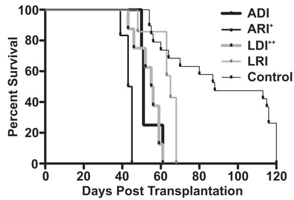
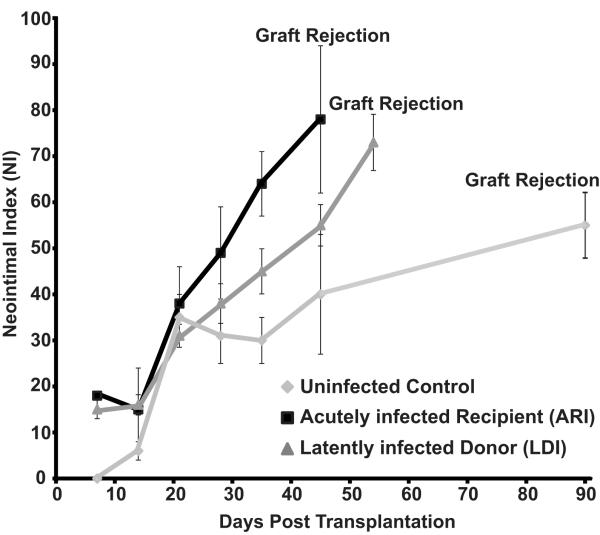
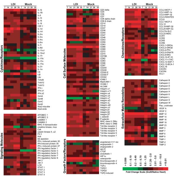
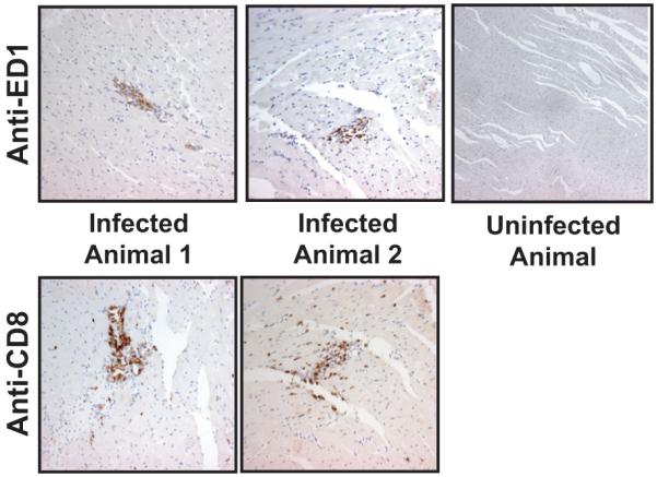
Human cytomegalovirus (HCMV) infection is associated with the acceleration of transplant vascular sclerosis (TVS) and chronic allograft rejection (CR). HCMV-negative recipients of latently HCMV infected donor grafts are at highest risk for developing CMV disease. Using a rat heart transplant CR model, we have previously shown that acute rat CMV (RCMV) infection following transplantation significantly accelerates both TVS and CR. Here, we report that RCMV-naïve recipients of heart allografts from latently RCMV-infected donors undergo acceleration of CR with similar kinetics as acutely infected recipients. In contrast to acutely infected recipients, treatment of recipients of latently infected donor hearts with ganciclovir did not prevent CR or TVS. We observed the formation of tertiary lymphoid structures (TLOs) containing macrophages and T cells in latently infected hearts prior to transplantation but not in uninfected rats. Moreover, pathway analysis of gene expression data from allografts from latently infected donors indicated an early and sustained production of TLO-associated genes compared to allografts from uninfected donors. We conclude that RCMV-induced TLO formation and alteration of donor tissue T cell profiles prior to transplantation in part mediate the ganciclovir-insensitive rejection of latently infected donor allografts transplanted into naïve recipients by providing a scaffold for immune activation.


Figures

Figure 1
Figure 1. RCMV infection source (donor vs. recipient) and status (acute and latent) are important for virus acceleration of heart allograft chronic rejection
Shown is a survival curve for heterotopic heart grafts (F344) transplanted into Lewis recipients and monitored over a 120-day period for clinical rejection. Groups included: 1. ADI (POD 51, donor was infected 7 days prior to transplantation, n=4); 2. ARI (POD 43, recipient was infected POD 1, n=6); 3. LDI (POD 55, donor was infected 180 days prior to transplantation, n=8); 4. LRI (POD 63, recipient was infected 180 prior to transplantation, n=7); and 5. Control (uninfected donor and recipient). + = ARI vs. ADI p<0.01, ++ = LDI vs. LRI p<0.01, ADI/ARI/LDI/LRI accelerate graft rejection vs. control p<0.001.
Figure 2
Figure 2. RCMV infection accelerates the time to develop TVS in graft hearts
Comparison of the time to allograft rejection and TVS (mean NI) of graft heart vessels of uninfected controls, RCMV-acutely infected recipients (ARI) and recipients of RCMV-latently infected donor hearts (LDI); n=6 for each. The time to rejection for the ARI and LDI was significantly decreased compared to that of the uninfected controls (p<0.05). Similarly, the increase NI parallels that of the time to rejection (p>0.05).
Figure 3
Figure 3. RCMV detection in salivary glands from recipients of latently infected donor hearts
RCMV genome copy was determined by real time PCR at 7, 14, 21, and 28 days post transplantation in recipients of latently infected donor hearts (n=4 at each time point).
Figure 4
Figure 4. Recipients of RCMV-latently infected donor hearts have predominately an IgM RCMV-specific Antibody Response
RCMV-specific ELISA plates were coated with lysates from RCMV infected fibroblasts. Plasma samples from allograft recipients of hearts from latently infected rats (LDI) at POD21, latently infected heart donor rats, acutely infected rats (21dpi) and uninfected controls (n=4 for each). Plasma was diluted 1:50. The secondary anti-rat IgM or IgG antibody conjugated to HRP was used for detection. Recipients of RCMV infected donor hearts have predominantly an IgM antibody response, which is statistically significant compared the IgG response (p<0.05).
Figure 5
Figure 5. Gene expression profiling identifies differences between allografts from recipients of latently infected donors and uninfected controls
Microarrays were prepared from total graft and native hearts from recipients of latently infected grafts (LDI) and uninfected controls (Mock) harvested at 7, 14, 21, 28, and 35-post transplantation. Shown is a heat map representing the fold change values of the allograft (n=3) compared to uninfected non-transplanted heart (n=4). The data shown is limited to the identified genes displaying significant dysregulation (up or down) when compared to control hearts (LDI vs. mock infected p<0.05 for at least one of the time points). The data were subdivided into categories based upon predicted biological function using the computer program GeneGo by Metagene.
Figure 6
Figure 6. RCMV Infection Induces SMC and Fibroblast Expression of TLO Associated Chemokines
Quantitative RT-PCR was used to quantify CCL19 (A), CCL21b (B), and CXCL13 (C) chemokine expression in samples of RCMV infected primary fibroblasts and aortic SMC. RNA samples were analyzed in triplicate and normalized to expression of L32 (cellular ribosomal protein), and the relative copy number was determined using amplicon standards to each gene.
Figure 7
Figure 7. Hearts from RCMV latently infected rats contain macrophages and T cells
Paraffin embedded heart sections from latently infected rats or controls were probed using antibodies directed against ED1 (Mac) and CD8 (T-cell). (mag=40x).
Figure 8
Figure 8. Latently infected rat hearts contain activated CD4 and CD8 T cells
Peripheral blood was harvested from uninfected rats or rats infected with RCMV at either 21 or 180 dpi (n=4 for each). Total lymphocytes were isolated from the rat hearts and these cells were stained for the cell surface markers CD4, CD8, and CD62 as well as for the intracellular marker of cellular proliferation, Ki67 and then analyzed by flow cytometry. Hearts from latently infected rats contain a significantly increased level of activated CD4+ and CD8+ effector T-cells compared to hearts from uninfected controls (p<0.05).

References

    1. Almond PS, Matas A, Gillingham K, Dunn DL, Payne WD, Gores P, Gruessner R, Najarian JS. Risk factors for chronic rejection in renal allograft recipients. Transplant. 1993;55(4):752–6. discussion 756-7. - PubMed
    1. Isoniemi H, Nurminen M, Tikkanen MJ, von Willebrand E, Krogerus L, Ahonen J, Eklund B, Hockerstedt K, Salmela K, Hayry P. Risk factors predicting chronic rejection of renal allografts. Transplantation. 1994;57(1):68–72. - PubMed
    1. Fitzgerald JT, Gallay B, Taranto SE, McVicar JP, Troppmann C, Chen X, McIntosh MJ, Perez RV. Pretransplant recipient cytomegalovirus seropositivity and hemodialysis are associated with decreased renal allograft and patient survival. Transplantation. 2004;77(9):1405–11. - PubMed
    1. Gnann JW, Jr., Ahlmen J, Svalander C, Olding L, Oldstone MB, Nelson JA. Inflammatory cells in transplanted kidneys are infected by human cytomegalovirus. Am J Pathol. 1988;132(2):239–48. - PMC - PubMed
    1. Grattan MT, Moreno-Cabral CE, Starnes VA, Oyer PE, Stinson EB, Shumway NE. Cytomegalovirus infection is associated with cardiac allograft rejection and atherosclerosis. JAMA. 1989;261(24):3561–6. - PubMed

Publication types