Infection of Arabidopsis thaliana by Phytophthora parasitica and identification of variation in host specificity
- PMID: 21199568
- PMCID: PMC6640465
- DOI: 10.1111/j.1364-3703.2010.00659.x
Infection of Arabidopsis thaliana by Phytophthora parasitica and identification of variation in host specificity
Abstract
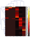
Oomycete pathogens cause severe damage to a wide range of agriculturally important crops and natural ecosystems. They represent a unique group of plant pathogens that are evolutionarily distant from true fungi. In this study, we established a new plant-oomycete pathosystem in which the broad host range pathogen Phytophthora parasitica was demonstrated to be capable of interacting compatibly with the model plant Arabidopsis thaliana. Water-soaked lesions developed on leaves within 3 days and numerous sporangia formed within 5 days post-inoculation of P. parasitica zoospores. Cytological characterization showed that P. parasitica developed appressoria-like swellings and penetrated epidermal cells directly and preferably at the junction between anticlinal host cell walls. Multiple haustoria-like structures formed in both epidermal cells and mesophyll cells 1 day post-inoculation of zoospores. Pathogenicity assays of 25 A. thaliana ecotypes with six P. parasitica strains indicated the presence of a natural variation in host specificity between A. thaliana and P. parasitica. Most ecotypes were highly susceptible to P. parasitica strains Pp014, Pp016 and Pp025, but resistant to strains Pp008 and Pp009, with the frequent appearance of cell wall deposition and active defence response-based cell necrosis. Gene expression and comparative transcriptomic analysis further confirmed the compatible interaction by the identification of up-regulated genes in A. thaliana which were characteristic of biotic stress. The established A. thaliana-P. parasitica pathosystem expands the model systems investigating oomycete-plant interactions, and will facilitate a full understanding of Phytophthora biology and pathology.
© 2010 Northwest A&F University. Molecular Plant Pathology © 2010 BSPP and Blackwell Publishing Ltd.
Figures


Similar articles
-
Phytophthora parasitica: a model oomycete plant pathogen.Mycology. 2014 Jun;5(2):43-51. doi: 10.1080/21501203.2014.917734. Epub 2014 May 19. Mycology. 2014. PMID: 24999436 Free PMC article. Review.
-
The immediate activation of defense responses in Arabidopsis roots is not sufficient to prevent Phytophthora parasitica infection.New Phytol. 2010 Jul;187(2):449-460. doi: 10.1111/j.1469-8137.2010.03272.x. Epub 2010 Apr 29. New Phytol. 2010. PMID: 20456058
-
Production of dsRNA sequences in the host plant is not sufficient to initiate gene silencing in the colonizing oomycete pathogen Phytophthora parasitica.PLoS One. 2011;6(11):e28114. doi: 10.1371/journal.pone.0028114. Epub 2011 Nov 23. PLoS One. 2011. PMID: 22140518 Free PMC article.
-
The Arabidopsis thaliana gene AtERF019 negatively regulates plant resistance to Phytophthora parasitica by suppressing PAMP-triggered immunity.Mol Plant Pathol. 2020 Sep;21(9):1179-1193. doi: 10.1111/mpp.12971. Epub 2020 Jul 28. Mol Plant Pathol. 2020. PMID: 32725756 Free PMC article.
-
Strategies of attack and defense in plant-oomycete interactions, accentuated for Phytophthora parasitica Dastur (syn. P. Nicotianae Breda de Haan).J Plant Physiol. 2008 Jan;165(1):83-94. doi: 10.1016/j.jplph.2007.06.011. Epub 2007 Sep 4. J Plant Physiol. 2008. PMID: 17766006 Review.
Cited by
-
Arabidopsis thaliana defense response to the ochratoxin A-producing strain (Aspergillus ochraceus 3.4412).Plant Cell Rep. 2015 May;34(5):705-19. doi: 10.1007/s00299-014-1731-3. Epub 2015 Feb 10. Plant Cell Rep. 2015. PMID: 25666274
-
Mutations in PpAGO3 Lead to Enhanced Virulence of Phytophthora parasitica by Activation of 25-26 nt sRNA-Associated Effector Genes.Front Microbiol. 2022 Mar 24;13:856106. doi: 10.3389/fmicb.2022.856106. eCollection 2022. Front Microbiol. 2022. PMID: 35401482 Free PMC article.
-
Pathogen-associated molecular pattern-triggered immunity and resistance to the root pathogen Phytophthora parasitica in Arabidopsis.J Exp Bot. 2013 Sep;64(12):3615-25. doi: 10.1093/jxb/ert195. Epub 2013 Jul 12. J Exp Bot. 2013. PMID: 23851194 Free PMC article.
-
Leucine-rich repeat receptor-like gene screen reveals that Nicotiana RXEG1 regulates glycoside hydrolase 12 MAMP detection.Nat Commun. 2018 Feb 9;9(1):594. doi: 10.1038/s41467-018-03010-8. Nat Commun. 2018. PMID: 29426870 Free PMC article.
-
Phytophthora parasitica: a model oomycete plant pathogen.Mycology. 2014 Jun;5(2):43-51. doi: 10.1080/21501203.2014.917734. Epub 2014 May 19. Mycology. 2014. PMID: 24999436 Free PMC article. Review.
References
-
- Adam, L. and Somerville, S.C. (1996) Genetic characterization of five powdery mildew disease resistance loci in Arabidopsis thaliana . Plant J. 9, 341–356. - PubMed
-
- Attard, A. , Gourgues, M. , Callemeyn‐Torre, N. and Keller, H. (2010) The immediate activation of defense responses in Arabidopsis roots is not sufficient to prevent Phytophthora parasitica infection. New Phytol. 187, 449–460. - PubMed
-
- van Baarlen, P. , Woltering, E.J. , Staats, M. and van Kan, J.A.L. (2007) Histochemical and genetic analysis of host and non‐host interactions of Arabidopsis with three Botrytis species: an important role for cell death control. Mol. Plant Pathol. 8, 41–54. - PubMed
-
- Barr, D.J.S. (1992) Evolution and kingdoms of organisms from the perspective of a mycologist. Mycologia, 84, 1–11.
-
- Bittner‐Eddy, P.D. and Beynon, J.L. (2001) The Arabidopsis downy mildew resistance gene, RPP13‐Nd, functions independently of NDR1 and EDS1 and does not require the accumulation of salicylic acid. Mol. Plant–Microbe Interact. 14, 416–421. - PubMed
Publication types
MeSH terms
LinkOut - more resources
Full Text Sources

