Review
. 2011 Jun 11;660(1):103-10.
doi: 10.1016/j.ejphar.2010.11.040. Epub 2011 Jan 1.

Melanocortin MC₁ receptor in human genetics and model systems

Affiliations
Review

Melanocortin MC₁ receptor in human genetics and model systems

Kimberley A Beaumont et al. Eur J Pharmacol. .

Abstract

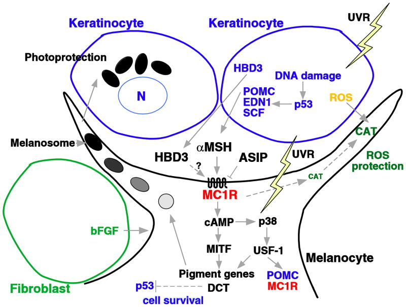
The melanocortin MC(1) receptor is a G-protein coupled receptor expressed in the melanocytes of the skin and hair and is known for its key role in the regulation of human pigmentation. Melanocortin MC(1) receptor activation after ultraviolet radiation exposure results in a switch from the red/yellow pheomelanin to the brown/black eumelanin pigment synthesis within cutaneous melanocytes; this pigment is then transferred to the surrounding keratinocytes of the skin. The increase in melanin maturation and uptake results in tanning of the skin, providing a physical protection of skin cells from ultraviolet radiation induced DNA damage. Melanocortin MC(1) receptor polymorphism is widespread within the Caucasian population and some variant alleles are associated with red hair colour, fair skin, poor tanning and increased risk of skin cancer. Here we will discuss the use of mouse coat colour models, human genetic association studies, and in vitro cell culture studies to determine the complex functions of the melanocortin MC(1) receptor and the molecular mechanisms underlying the association between melanocortin MC(1) receptor variant alleles and the red hair colour phenotype. Recent research indicates that melanocortin MC(1) receptor has many non-pigmentary functions, and that the increased risk of skin cancer conferred by melanocortin MC(1) receptor variant alleles is to some extent independent of pigmentation phenotypes. The use of new transgenic mouse models, the study of novel melanocortin MC(1) receptor response genes and the use of more advanced human skin models such as 3D skin reconstruction may provide key elements in understanding the pharmacogenetics of human melanocortin MC(1) receptor polymorphism.


Figures

Figure 1
Figure 1
The melanocyte and the tanning response. Adapted from (Beaumont et al., 2009). Exposure of human skin to ultraviolet radiation results in the release of various factors by keratinocytes and melanocytes. Proopiomelanocortin (POMC) is converted to α-melanocyte stimulating hormone ( MSH), which stimulates melanocortin MC1 receptor (α-MC1R) cAMP signaling in the melanocyte causing the upregulation of genes involved in melanogenesis, ultimately resulting in the tanning response. Agouti signaling protein (ASIP) anatgonises the action of α-MSH. Dotted lines and ? indicate interactions that are in need of further research. ROS, reactive oxygen species; UVR = ultraviolet radiation, HBD3 = human β-defensin 3, END1 = endothelin 1, SCF, stem cell factor; CAT, catalase; bFGF, basic fibroblast growth factor; MITF, microphthalmia transcription factor; DCT, dopachrome tautomerase; USF-1, upstream transcription factor 1.
Figure 2
Figure 2
cAMP accumulation in B16 or HEK293 cells stably transfected with melanocortin MC1 receptor (HEKWTe). Cells were incubated in serum-free media for at least 2 hours. Cells were pre-incubated with 0.1mM IBMX for 15min, then stimulated with the indicated ligands for 10min. cAMP levels were quantified with the cAMP EIA system (Amersham Biosciences). The bars indicate the range from two independent experiments A) 0.5nM or 1nM NDP-MSH alone or in combination with 100nM agouti signaling protein peptide (ASIP-YY) (McNulty et al., 2005) or HBD3, or 100nM ASIP-YY or HBD3 alone. Data was normalised to NDP-MSH, which was set at 100%. Results are from two independent experiments. B) 1nM NDP-MSH, 100nM HBD3 or 100nM ASIP-YY. Data was expressed as fold over the control. Results are from 2 independent experiments, except for NDP-MSH, which is from only one experiment.
Figure 3
Figure 3
ERK phosphorylation in response to α-melanocyte stimulating hormone and human β-Defensin 3. Lanes are as indicated C=Control, N=10nM NDP-MSH, β=10nM HBD3. HEK293 untransfected cells (parental) or HEK293 stably expressing melanocortin MC 1 receptor (WTe) were pre-incubated in serum-free media for at least 2 hours, then stimulated with the indicated ligands for 5min. Western immuno-blotting with the indicated antibodies was performed using total cell extracts. pERK (Cell Signalling) is specific to the phosphorylated Thr202/Tyr204 sites of ERK1/2, ERK (Cell Signalling) recognises all forms of the ERK protein. Anti-GAPDH (R&D Systems) was used as a loading control. This blot is representative of three independent experiments.
Figure 4
Figure 4
Melanocyte co-culture p38 and p53 responses to NDP-MSH and ultraviolet radiation. Co-cultures of melanocortin MC1 receptor wild type (WT–QF1193) or homozygous R151C melanocytes (QF1108) and keratinocytes were treated with 20 mJ/cm2 ultraviolet radiation(U), 20 nM NDP -MSH (N) or both (UN) and incubated for 1 hr. Total cell extracts were used for western immuno-blotting. Activated forms of p38 and p53 were detected using antibodies specific for the Thr180/Tyr182 (pp38) and Ser15 (pp53) phosphorylated sites (Cell Signaling). These blots are representative of 2-3 independent experiments.
Figure 5
Figure 5
Dopachrome tautomeraseinduction in melanocyte co -cultures. Melanocytes were either melanocortin MC1 receptor wild type (WT–QF1106 strain), heterozygous for R151C (QF1200 strain) or heterozygous for R160W (QF1100 strain). Co-cultures of melanocytes with keratinocytes were treated with the following compounds: C = Control, N = 20nM NDP-MSH or F = 10uM forskolin, for four days, then total cell extracts were used for western immuno-blotting with dopachrome tautomerase(Santa Cruz) and GAPDH antibodies. This blot is representative of responses seen in three different wild type and variant strains.

References

    1. Abdel-Malek ZA, Kadekaro AL, Swope VB. Stepping up melanocytes to the challenge of UV exposure. Pigment Cell Melanoma Res. 2010;23:171–186. - PubMed
    1. Abdel-Malek ZA, Knittel J, Kadekaro AL, Swope VB, Starner R. The melanocortin 1 receptor and the UV response of human melanocytes--a shift in paradigm. Photochem Photobiol. 2008;84:501–508. - PubMed
    1. Bastiaens MT, ter Huurne JAC, Kielich C, Gruis NA, Westendorp RGJ, Vermeer BJ, Bavinck NJB. Melanocortin-1 receptor gene variants determine the risk of nonmelanoma skin cancer independently of fair skin and red hair. American Journal of Human Genetics. 2001;68:884–894. - PMC - PubMed
    1. Beaumont KA, Liu YY, Sturm RA. Chapter 4 The Melanocortin-1 Receptor Gene Polymorphism and Association with Human Skin Cancer. Prog Mol Biol Transl Sci. 2009;88C:85–153. - PubMed
    1. Beaumont KA, Newton RA, Smit DJ, Leonard JH, Stow JL, Sturm RA. Altered cell surface expression of human MC1R variant receptor alleles associated with red hair and skin cancer risk. Hum Mol Genet. 2005;14:2145–2154. - PubMed

Publication types

Substances

LinkOut - more resources