. 2011 Mar 15;20(6):1154-63.
doi: 10.1093/hmg/ddq559. Epub 2011 Jan 3.

Over-expression of BCL2 rescues muscle weakness in a mouse model of oculopharyngeal muscular dystrophy

Affiliations

Over-expression of BCL2 rescues muscle weakness in a mouse model of oculopharyngeal muscular dystrophy

Janet E Davies et al. Hum Mol Genet. .

Abstract

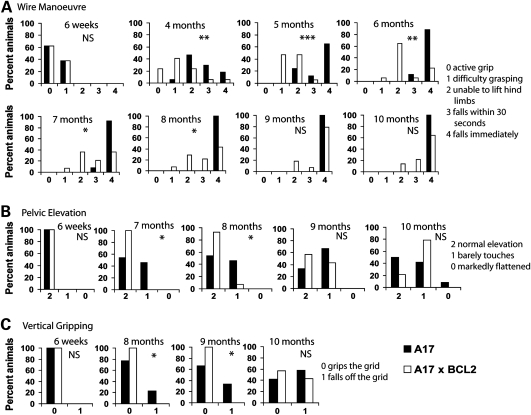
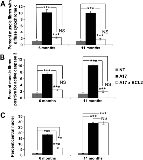
Oculopharyngeal muscular dystrophy (OPMD) is a late-onset muscular dystrophy caused by a polyalanine expansion mutation in the coding region of the poly-(A) binding protein nuclear 1 (PABPN1) gene. In unaffected individuals, (GCG)(6) encodes the first 6 alanines in a homopolymeric stretch of 10 alanines. In most patients, this (GCG)(6) repeat is expanded to (GCG)(8-13), leading to a stretch of 12-17 alanines in mutant PABPN1, which is thought to confer a toxic gain of function. Thus, OPMD has been modelled by expressing mutant PABPN1 transgenes in the presence of endogenous copies of the gene in cells and mice. In these models, increased apoptosis is seen, but it is unclear whether this process mediates OPMD. The role of apoptosis in the pathogenesis of different muscular dystrophies is unclear. Blocking apoptosis ameliorates muscle disease in some mouse models of muscular dystrophy such as laminin α-2-deficient mice, but not in others such as dystrophin-deficient (mdx) mice. Here we demonstrate that apoptosis is not only involved in the pathology of OPMD but also is a major contributor to the muscle weakness and dysfunction in this disease. Genetically blocking apoptosis by over-expressing BCL2 ameliorates muscle weakness in our mouse model of OPMD (A17 mice). The effect of BCL2 co-expression on muscle weakness is transient, since muscle weakness is apparent in mice expressing both A17 and BCL2 transgenes at late time points. Thus, while apoptosis is a major pathway that causes muscle weakness in OPMD, other cell death pathways may also contribute to the disease when apoptosis is inhibited.


Figures

Figure 1.
Figure 1.
BCL2 expression improves grip strength at early time points in a mouse model of OPMD. (A) Western blot of biceps muscle lysates from 6 months and 11 months, A17 mice, non-transgenic mice (NT), BCL2 mice and A17 × BCL2 mice probed with antibodies against PABPN1 or human BCL2 (to show transgene levels). Tubulin was used as a loading control. (B and C) Forelimb grip strength (B) and grip strength of all limbs (C) of male A17 mice (black triangle; 6 weeks, n = 17; 4, 5 and 6 months, n = 17; 7 and 8 months, n = 13, 9 and 10 months, n = 12), non-transgenic mice (NT; grey diamond; 6 weeks, n = 14; 4, 5 and 6 months, n = 14; 7 and 8 months, n = 11; 9 and 10 months, n = 10), BCL2 mice (black square; 6 weeks, n = 17; 4, 5 and 6 months, n = 17; 7 and 8 months, n = 13; 9 and 10 months, n = 12) and mice expressing both A17 and BCL2 transgenes (A17 × BCL2; open circle; 6 weeks, n = 17; 4, 5 and 6 months, n = 17; 7, 8, 9 and 10 months, n = 14). (B) Forelimb grip strength was improved in A17 × BCL2 mice compared with A17 mice at 4 months (P < 0.0001), 5 months (P < 0.0001), 6 months (P < 0.0001), 7 months (P < 0.0001) and 8 months (P = 0.0003) of age but not at 9 months (P = 0.08) and 10 months (P = 0.06) of age. Repeated measure ANOVAs for determination of the overall effect from all time points P, <0.0001. (C) Grip strength from all limbs was improved in A17 × BCL2 mice compared with A17 mice at 4 months (P < 0.0001), 5 months (P < 0.0001), 6 months (P < 0.0001), 7 months (P < 0.0001) and 8 months (P = 0.0006) of age but not at 9 months (P = 0.06) and 10 months (P = 0.3) of age. Repeated measure ANOVAs for determination of the overall effect from all time points P, <0.0001. No significant difference in forelimb grip strength or grip strength from all limbs was seen between BCL2 mice and non-transgenic mice at all time points studied, or between any of the different groups at 6 weeks of age. Data were analysed using two-tailed Student's t-tests at each individual time point. Error bars represent SD.
Figure 2.
Figure 2.
BCL2 expression transiently improves muscle strength in a mouse model of OPMD. Male A17 mice (black bar; 6 weeks, n = 17; 4, 5 and 6 months, n = 17; 7 and 8 months, n = 13; 9 and 10 months, n = 12) and male A17 × BCL2 mice (open bar; 6 weeks, n = 17; 4, 5 and 6 months, n = 17; 7, 8, 9 and 10 months, n = 14) were compared in strength tests. (A) A17 × BCL2 mice performed better than A17 mice at the wire manoeuvre task at 4, 5, 6, 7 and 8 months of age but not at 9 and 10 months of age. Score: 0, active grip with hind legs; 1, difficulty grasping with hind legs; 2, unable to lift hind legs; 3, falls within 30 s; 4, falls immediately. (B) Pelvic elevation was improved in A17 × BCL2 mice compared with A17 mice at the ages of 7 and 8 months but not at 9 and 10 months. Score: 2, normal elevation; 1, barely touches; 0, markedly flattened. (C) A17 × BCL2 males performed significantly better at the vertical gripping test at 8 and 9 months of age, but not at 10 months of age, compared with A17 males. Score: 0, grips the grid; 1, falls off the grid. *P < 0.05; **P < 0.001; ***P < 0.0001; NS, non-significant. Non-parametric data from the wire manoeuvre and pelvic elevation tests were analysed using Mann–Whitney U-tests and we used Chi-squared tests to analyse vertical gripping data.
Figure 3.
Figure 3.
Weight differences between non-transgenic, A17, BCL2 and A17 × BCL2 mice. (A) Body weight of male A17 mice (black triangle; 6 weeks, n = 17; 4, 5 and 6 months, n = 17; 7 and 8 months, n = 13, 9 and 10 months, n = 12), non-transgenic mice (NT; grey diamond; 6 weeks, n = 14; 4, 5 and 6 months, n = 14; 7 and 8 months, n = 11; 9 and 10 months, n = 10), BCL2 mice (black square; 6 weeks, n = 17; 4, 5 and 6 months, n = 17; 7 and 8 months, n = 13; 9 and 10 months, n = 12) and mice expressing both A17 and BCL2 transgenes (A17 × BCL2; open circle; 6 weeks, n = 17; 4, 5 and 6 months, n = 17; 7, 8, 9 and 10 months, n = 14). Male A17 mice on a mixed FvB C57BL/6 background weighed less than non-transgenic littermates at 4 months (P = 0.0001), 5 months (P < 0.0001), 6 months (P = 0.0004), 7 months (P = 0.004), 8 months (P = 0.0008), 9 months (P = 0.002) and 10 months of age (P = 0.002). Repeated measure ANOVAs for determination of the overall effect from all time points P, <0.0001. Co-expression of BCL2 in A17 mice alters the distribution of body weights, and A17 × BCL2 mice generally weighed more than A17 mice at 4 months (P = 0.04), 5 months (P = 0.02), 6 months (PP = 0.04), 7 months (P = 0.009), 8 months (P = 0.03), 9 months (P = 0.03) and 10 months of age (P = 0.008) (repeated measure ANOVAs for determination of the overall effect from all time points P, <0.0001). The expression of BCL2 alone does not alter body weight, and there was no significant difference in body weight between BCL2 mice and NT mice at any time point studied. There was no difference in body weight between any of the groups at 6 weeks of age. (B) Muscle mass of quadriceps and tibilias anterior muscles from 6-month-old A17, NT, BCL2 and A17 × BCL2 mice (n = 3). (C) Muscle mass of quadriceps and tibilias anterior muscles from 11-month-old A17, NT, BCL2 and A17 × BCL2 mice (n = 3). *P < 0.05; **P < 0.001; ***P < 0.0001; NS, non-significant. Error bars represent SEM.
Figure 4.
Figure 4.
Cell death and aggregate load in A17 mice and mice expressing both A17 and BCL2 transgenes. (A) Biceps muscle sections from 6-month-old and 11-month-old A17 (black bar), NT (grey bar) and A17 × BCL2 (open bar) mice were TUNEL labelled and the number of positive nuclei scored. (B) Representative images of PABPN1-labelled aggregates (green) in biceps muscle sections from 6-month-old A17 mice and A17 × BCL2 mice. Sections were treated with KCl prior to labelling to remove soluble proteins (aggregates are resistant) and hence no signal is seen in sections from non-transgenic (NT) mice. Nuclei were visualized using DAPI (blue). (C) Quantification of the number of nuclei containing PABPN1-labelled aggregates in sections from 6-month-old and 11-month-old A17 mice (black bar) and A17 × BCL2 (open bar) mice treated as (B). n = 3; *P < 0.05; ***P < 0.0001. Error bars represent SEM.
Figure 5.
Figure 5.
Apoptotic markers are elevated in muscle from A17 mice and reduced by BCL2 co-expression. (A) Quantification of the number of myofibres with a diffuse pattern of cytochrome c labelling (corresponding to cytochrome c release from the mitochondria to cytosol and indicating apoptotic myofibres) in biceps muscle sections from 6-month-old and 12-month-old NT, A17 and A17 × BCL2 mice. (B) Biceps muscle sections from 6-month-old and 12-month-old NT, A17 and A17 × BCL2 mice were labelled with an active caspase 3 antibody and the number of immuno-positive myofibres were scored. (C) Quantification of the number of centralized nuclei in H&E stained biceps sections from 6-month-old and 12-month-old NT, A17 and A17 × BCL2 mice. Apoptotic markers are reduced in A17 × BCL2 mice to NT levels even at 11 months when BCL2 does not rescue phenotype and muscle weakness is apparent in A17 × BCL2 mice (n = 3). **P < 0.001; ***P < 0.0001; NS, non-significant. Error bars represent SEM.
Figure 6.
Figure 6.
A central role for apoptosis in the pathogenesis of OPMD. The expression of mutant PABPN1 (A17) results in increased levels of BAX (12). This causes the activation of the apoptotic cascade (12,15). Cytochrome c is released from the mitochondria to the cytosol where it can bind additional factors to form the apoptosome complex. The apoptosome complex causes the conversion of procaspases to their active, mature caspase form and ultimately results in the activation of caspase 3, otherwise known as the executioner caspase. Here we have provided data to further support a central role of apoptosis in the pathogenesis of OPMD. Genetically blocking apoptosis by the over-expression of BCL2 ameliorates A17 toxicity. BCL2 most likely acts to block A17 toxicity by binding the excess BAX and sequestering it away from the mitochondria. Wild-type PABPN1 has an opposing effect on apoptosis and increases the translation of XIAP, a caspase inhibitor (38). It is likely that OPMD is primarily caused by a toxic gain of function of mutant PABPN1 that results in the activation of apoptosis. However, the loss of the anti-apoptotic function of wild-type PABPN1 may also contribute to pathology.

Similar articles

Cited by

  • Systemic Delivery of a Monoclonal Antibody to Immunologically Block Myostatin in the A17 Mouse Model of OPMD.
    Malerba A, Harish P, Popplewell L. Malerba A, et al. Methods Mol Biol. 2023;2587:557-568. doi: 10.1007/978-1-0716-2772-3_30. Methods Mol Biol. 2023. PMID: 36401050
  • Apoptotic cell death in disease-Current understanding of the NCCD 2023.
    Vitale I, Pietrocola F, Guilbaud E, Aaronson SA, Abrams JM, Adam D, Agostini M, Agostinis P, Alnemri ES, Altucci L, Amelio I, Andrews DW, Aqeilan RI, Arama E, Baehrecke EH, Balachandran S, Bano D, Barlev NA, Bartek J, Bazan NG, Becker C, Bernassola F, Bertrand MJM, Bianchi ME, Blagosklonny MV, Blander JM, Blandino G, Blomgren K, Borner C, Bortner CD, Bove P, Boya P, Brenner C, Broz P, Brunner T, Damgaard RB, Calin GA, Campanella M, Candi E, Carbone M, Carmona-Gutierrez D, Cecconi F, Chan FK, Chen GQ, Chen Q, Chen YH, Cheng EH, Chipuk JE, Cidlowski JA, Ciechanover A, Ciliberto G, Conrad M, Cubillos-Ruiz JR, Czabotar PE, D'Angiolella V, Daugaard M, Dawson TM, Dawson VL, De Maria R, De Strooper B, Debatin KM, Deberardinis RJ, Degterev A, Del Sal G, Deshmukh M, Di Virgilio F, Diederich M, Dixon SJ, Dynlacht BD, El-Deiry WS, Elrod JW, Engeland K, Fimia GM, Galassi C, Ganini C, Garcia-Saez AJ, Garg AD, Garrido C, Gavathiotis E, Gerlic M, Ghosh S, Green DR, Greene LA, Gronemeyer H, Häcker G, Hajnóczky G, Hardwick JM, Haupt Y, He S, Heery DM, Hengartner MO, Hetz C, Hildeman DA, Ichijo H, Inoue S, Jäättelä M, Janic A, Joseph B, Jost PJ, Kanneganti TD, Karin M, Kashkar H, Kaufmann T, Kelly … See abstract for full author list ➔ Vitale I, et al. Cell Death Differ. 2023 May;30(5):1097-1154. doi: 10.1038/s41418-023-01153-w. Epub 2023 Apr 26. Cell Death Differ. 2023. PMID: 37100955 Free PMC article. Review.
  • Mitochondrial dysfunction reveals the role of mRNA poly(A) tail regulation in oculopharyngeal muscular dystrophy pathogenesis.
    Chartier A, Klein P, Pierson S, Barbezier N, Gidaro T, Casas F, Carberry S, Dowling P, Maynadier L, Bellec M, Oloko M, Jardel C, Moritz B, Dickson G, Mouly V, Ohlendieck K, Butler-Browne G, Trollet C, Simonelig M. Chartier A, et al. PLoS Genet. 2015 Mar 27;11(3):e1005092. doi: 10.1371/journal.pgen.1005092. eCollection 2015 Mar. PLoS Genet. 2015. PMID: 25816335 Free PMC article.
  • SIRT1-dependent myoprotective effects of resveratrol on muscle injury induced by compression.
    Sin TK, Yung BY, Yip SP, Chan LW, Wong CS, Tam EW, Siu PM. Sin TK, et al. Front Physiol. 2015 Oct 21;6:293. doi: 10.3389/fphys.2015.00293. eCollection 2015. Front Physiol. 2015. PMID: 26557094 Free PMC article.
  • A Systematic Review of Oropharyngeal Dysphagia Models in Rodents.
    Kim HN, Kim JY. Kim HN, et al. Int J Environ Res Public Health. 2021 May 7;18(9):4987. doi: 10.3390/ijerph18094987. Int J Environ Res Public Health. 2021. PMID: 34067192 Free PMC article.

References

    1. Abu-Baker A., Rouleau G.A. Oculopharyngeal muscular dystrophy: recent advances in the understanding of the molecular pathogenic mechanisms and treatment strategies. Biochim. Biophys. Acta. 2007;1772:173–185. - PubMed
    1. Davies J.E., Berger Z., Rubinsztein D.C. Oculopharyngeal muscular dystrophy: potential therapies for an aggregate-associated disorder. Int. J. Biochem. Cell Biol. 2006;38:1457–1462. doi:10.1016/j.biocel.2006.01.016. - DOI - PubMed
    1. Schmitt H.P., Krause K.H. An autopsy study of a familial oculopharyngeal muscular dystrophy (OPMD) with distal spread and neurogenic involvement. Muscle Nerve. 1981;4:296–305. doi:10.1002/mus.880040406. - DOI - PubMed
    1. van der Sluijs B.M., van Engelen B.G., Hoefsloot L.H. Oculopharyngeal muscular dystrophy (OPMD) due to a small duplication in the PABPN1 gene. Hum. Mutat. 2003;21:553. doi:10.1002/humu.9138. - DOI - PubMed
    1. Blumen S.C., Brais B., Korczyn A.D., Medinsky S., Chapman J., Asherov A., Nisipeanu P., Codere F., Bouchard J.P., Fardeau M., et al. Homozygotes for oculopharyngeal muscular dystrophy have a severe form of the disease. Ann. Neurol. 1999;46:115–118. doi:10.1002/1531-8249(199907)46:1&lt;115::AID-ANA17&gt;3.0.CO;2-O. - DOI - PubMed

Publication types

Substances