. 2011 Mar 11;286(10):8481-8492.
doi: 10.1074/jbc.M110.174516. Epub 2011 Jan 3.

Conserved intramolecular disulfide bond is critical to trafficking and fate of ATP-binding cassette (ABC) transporters ABCB6 and sulfonylurea receptor 1 (SUR1)/ABCC8

Affiliations

Conserved intramolecular disulfide bond is critical to trafficking and fate of ATP-binding cassette (ABC) transporters ABCB6 and sulfonylurea receptor 1 (SUR1)/ABCC8

Yu Fukuda et al. J Biol Chem. .

Abstract

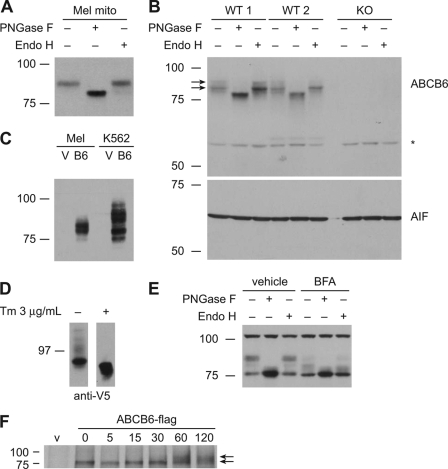
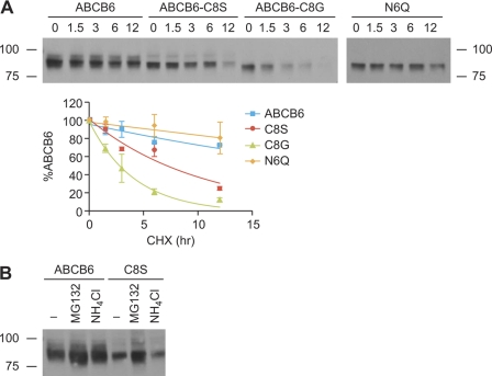
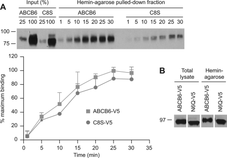
The ATP-binding cassette (ABC) transporter ABCB6 is a mitochondrial porphyrin transporter that activates porphyrin biosynthesis. ABCB6 lacks a canonical mitochondrial targeting sequence but reportedly traffics to other cellular compartments such as the plasma membrane. How ABCB6 reaches these destinations is unknown. In this study, we show that endogenous ABCB6 is glycosylated in multiple cell types, indicating trafficking through the endoplasmic reticulum (ER), and has only one atypical site for glycosylation (NXC) in its amino terminus. ABCB6 remained glycosylated when the highly conserved cysteine (Cys-8) was substituted with serine to make a consensus site, NXS. However, this substitution blocked ER exit and produced ABCB6 degradation, which was mostly reversed by the proteasomal inhibitor MG132. The amino terminus of ABCB6 has an additional highly conserved ER luminal cysteine (Cys-26). When Cys-26 was mutated alone or in combination with Cys-8, it also resulted in instability and ER retention. Further analysis revealed that these two cysteines form a disulfide bond. We discovered that other ABC transporters with an amino terminus in the ER had similarly configured conserved cysteines. This analysis led to the discovery of a disease-causing mutation in the sulfonylurea receptor 1 (SUR1)/ABCC8 from a patient with hyperinsulinemic hypoglycemia. The mutant allele only contains a mutation in a conserved amino-terminal cysteine, producing SUR1 that fails to reach the cell surface. These results suggest that for ABC transporters the propensity to form a disulfide bond in the ER defines a unique checkpoint that determines whether a protein is ER-retained.


Figures

FIGURE 1.
FIGURE 1.
ABCB6 is glycosylated and traffics through ER. Crude mitochondrial (mito) preparations from murine erythroleukemia (Mel) cells (A) and liver isolated from two wild-type (WT) mice and one Abcb6 knock-out (KO) mouse (B) were treated with PNGase F or Endo H, and ABCB6 was detected by immunoblotting using an anti-ABCB6 antibody. Immunoblotting was performed on one set of samples with biological duplicates. Apoptosis inducing factor (AIF) is shown as a control for equal loading of the proteins. An asterisk indicates nonspecific bands that appear both in WT and knock-out. C, analysis of hABCB6-FLAG in crude mitochondria preparations from Mel (mouse) and K562 (human) cells using an anti-FLAG antibody shows different glycosidation patterns. D, ABCB6 was no longer glycosylated when K562 cells expressing ABCB6-V5 were treated with tunicamycin. An immunoblot from a representative experiment is shown. E, crude mitochondrial preparations from Mel cells treated with vehicle (DMSO) or 1 μg/ml brefeldin A (BFA) for 24 h were analyzed for glycosidase sensitivity using an anti-ABCB6 antibody. F, pulse-chase assay using K562 cells expressing ABCB6-FLAG showed the glycosylated ABCB6 at 60 min. Tm, tunicamycin. Images are from at least two independent experiments unless otherwise noted.
FIGURE 2.
FIGURE 2.
ABCB6 is modified at single conserved atypical glycosylation site. A, ABCB6 contains four consensus N-glycosylation (NX(S/T)) motifs and a single atypical N-glycosylation motif. B, ABCB6 with amino acid substitution of all four asparagines in the consensus motif (Q4) was analyzed for PNGase F and Endo H sensitivity by immunoblotting using anti-V5 antibody. The presence of glycans was detected by protein mobility shifts. The migration patterns of all mutants were similar to those of the wild-type protein. C, NIH3T3 cells were transiently transfected with single mutant N6Q, Q4, or Q5 in which glutamine was substituted for all five asparagine residues. Cell lysates were treated with the indicated glycosidases and immunoblotted with anti-V5 antibody (lower arrow, non-glycosylated ABCB6; upper arrow, glycosylated ABCB6). D, multiple sequence alignment of ABCB6 shows two conserved cysteines (red boxes) and one N-glycosylation site (green box) among the species. Each experimental result was independently performed in all iterations shown at least twice with a single representative blot from one experiment used. vec, vector; H.sapiens, Homo sapiens; P.troglodytes, Pan troglodytes; B.taurus, Bos taurus; M.musculus, Mus musculus; R.norvegicus, Rattus norvegicus; D.rerio, Danio rerio. Pro, protein (amino acid) sequence.
FIGURE 3.
FIGURE 3.
Cys-8 is dispensable for glycosylation but is unstable. A, Cys-8 was substituted with serine or glycine, and the mutant proteins were transiently expressed in NIH3T3 cells. The glycan modification of the mutants was determined by PNGase F sensitivity and analyzed by immunoblotting using anti-V5 antibody. ABCB6 was still glycosylated when Cys-8 was substituted with Ser to make a consensus N-glycosylation motif. B, K562 cells were transduced with plasmids containing IRES-GFP and wild-type ABCB6-, Walker A mutant (mt)-, C8S-, or C26A-FLAG. Cells were sorted for GFP fluorescence by FACS and analyzed by immunoblotting using anti-FLAG antibody for the expression of ABCB6 proteins. Cysteine mutants C8S and C26A were expressed at low levels. An immunoblot from a single experiment is shown. C, serine was substituted for Cys-8 and/or Cys-26 in ABCB6, or a cysteine residue was inserted between positions 9 and 10 in the C8S mutant (C8S/C10in). Endo H sensitivity indicates that all cysteine mutants failed to exit the ER. Cys-10 insertion did not restore the protein expression level nor the impaired ER exit. D, K562 cells stably expressing wild-type ABCB6, C8S, or C26A were labeled with [35S]Met/Cys for 5 min, washed, and chased for the indicated times. Cells were harvested, and immunoprecipitated FLAG-tagged proteins were separated by SDS-PAGE and detected by phosphorimaging. The amount of ABCB6 proteins was analyzed by densitometry and plotted as percentage of ABCB6 protein at 0 h for each construct. Values shown are mean with the S.D. indicated by the error bars from three independent experiments. Unless otherwise noted, all experiments were repeated at least twice. A representative image from one complete experiment is shown.
FIGURE 4.
FIGURE 4.
Glycosylation is not critical for ABCB6 instability, and MG132 partially rescues C8S instability. A, NIH3T3 cells were transfected with ABCB6-V5, C8S-V5, C8G-V5, or N6Q-V5, and cycloheximide (final concentration, 50 μg/ml) was added 17 h post-transfection. Cells were harvested at the indicated times and analyzed for ABCB6 proteins using anti-V5 antibody. Intensity of the bands was analyzed using densitometry and expressed as percentage of ABCB6 protein at 0 h for each construct. Curve fitting was performed by non-linear regression analysis using GraphPad Prism. Values shown are the mean from two independent experiments with the range indicated by the error bars. B, NIH3T3 cells were transiently transfected with ABCB6-V5 or C8S-V5, incubated with 10 μm MG132 or 25 mm NH4Cl for 12 h, and analyzed for ABCB6 proteins using an anti-V5 antibody. CHX, cycloheximide. A representative image from two separate experiments is shown.
FIGURE 5.
FIGURE 5.
Heme binding by ABCB6 does not require Cys-8 or glycosylation. A, ABCB6-V5 or ABCB6-C8S-V5 was transiently expressed in NIH3T3 cells. The cell lysates were incubated with hemin-agarose for the indicated periods (min), and the beads were washed. ABCB6 protein bound to the hemin-agarose was analyzed by immunoblot. The intensity of the immunoblot bands was analyzed by densitometry and is shown as percentage of maximum binding. Values shown are the mean from two independent experiments with the range indicated by the error bars. B, ABCB6-V5 or N6Q-V5 was transiently expressed in NIH3T3 cells, and the lysates were subjected to hemin-agarose pulldown assays. An immunoblot from a single experiment that was replicated many times (13) is shown.
FIGURE 6.
FIGURE 6.
ABCB6 amino-terminal cysteines form a disulfide bond. A, an ABCB6-FLAG chimera including amino acids N1–210 is predicted by TMHMM to contain five TM helices. The diagram is not drawn to scale. The glycosylation site (Asn) and two cysteines are labeled. B, ABCB6-C50A/C120AN1–210-FLAG and ABCB6-C8S/C50A/C120AN1–210-FLAG were expressed in NIH3T3 cells, treated with N-ethylmaleimide to prevent spontaneous disulfide bonds, separated by SDS-PAGE with or without DTT, and analyzed by immunoblotting. The identity of each band was confirmed by glycosidase sensitivity. Band I, mature ABCB6; band II, immature ABCB6 with high mannose modification; band III, nonglycosylated ABCB6. Asterisks indicate oxidized ABCB6 (faster migration in the absence of DTT). The right panel depicts a longer film exposure; however, all samples were analyzed on a single gel (blot). Representative images from two separate experiments are shown. C, intracellular PPIX content in K562 cells transduced with GFP and with ABCB6-FLAG, the Walker A mutant (mt)-FLAG, or ABCB6-C26A-FLAG was measured by flow cytometric analysis of GFP-positive cells. The values shown are mean with the S.D. indicated by the error bars from three independent experiments. Representative histograms are shown with a representative FACS dot plot that was used to generate these data depicted in the supplemental material.
FIGURE 7.
FIGURE 7.
Two amino-terminal cysteine residues are conserved and functional in ABCC transporters. A, a point mutation resulting in Cys-6 to Ser substitution in SUR1/ABCC8 was identified in one allele of a hyperinsulinemic patient as shown in the electropherogram. B, SUR1 and mutant SUR1 constructs containing various cysteine substitutions were co-expressed with KIR6.2 in COSm6 cells. SUR1 proteins were labeled with 125I-azidoglibenclamide and detected by autoradiography after SDS-PAGE. Mature SUR1 that traffics to the cell surface was identified by its slower electrophoretic migration. C, multiple sequence alignment of SUR1 and ABCC1/MRP1 show that the amino-terminal cysteine residues (boxed in red) are conserved among the species. D, MRP1-MSD0-GFP containing either wild-type MRP1 or a C32A-MRP1 mutant was transiently expressed in HEK293 cells. Merged indirect immunofluorescence microscopy images (left) show the wild-type protein (green) on the cell surface, whereas the C32A mutant protein remains in the ER where it co-localizes with the ER protein protein-disulfide isomerase (PDI) (red, which is shown as yellow in the co-localized cell). Scale bar, 10 μm. Proteins were treated with the indicated glycosidases and analyzed by immunoblotting with anti-GFP antibody (right). The presence of glycans was detected by protein mobility shifts. Two independent experiments were performed with all iterations. Representative images of a complete experiment are shown. E, cysteine residues between TM5 and TM6 of ABCG2 that form an intramolecular disulfide bond are boxed in red. A cysteine residue that participates in intermolecular disulfide bond formation is boxed in blue. ABCG5 also contains two cysteine residues in a similar region. H.sapiens, Homo sapiens; B.taurus, Bos taurus; M.musculus, Mus musculus; R.norvegicus, Rattus norvegicus; D.rerio, Danio rerio; G.gallus; Gallus gallus; C.lupus; Canis lupus. Pro, protein (amino acid) sequence.

Similar articles

Cited by

References

    1. Dean M. (2005) Methods Enzymol. 400, 409–429 - PubMed
    1. Riordan J. R., Rommens J. M., Kerem B., Alon N., Rozmahel R., Grzelczak Z., Zielenski J., Lok S., Plavsic N., Chou J. L., Drumm M. L., Iannuzzi M. C., Collins F. S., Tsui L. (1989) Science 245, 1066–1073 - PubMed
    1. Cheng S. H., Gregory R. J., Marshall J., Paul S., Souza D. W., White G. A., O'Riordan C. R., Smith A. E. (1990) Cell 63, 827–834 - PubMed
    1. Ellgaard L., Molinari M., Helenius A. (1999) Science 286, 1882–1888 - PubMed
    1. Ellgaard L., Helenius A. (2003) Nat. Rev. Mol. Cell Biol. 4, 181–191 - PubMed

Publication types

MeSH terms

LinkOut - more resources