. 2011 Mar 4;286(9):7609-18.
doi: 10.1074/jbc.M110.166603. Epub 2011 Jan 3.

Inhibition of MAPK by prolactin signaling through the short form of its receptor in the ovary and decidua: involvement of a novel phosphatase

Affiliations

Inhibition of MAPK by prolactin signaling through the short form of its receptor in the ovary and decidua: involvement of a novel phosphatase

Y Sangeeta Devi et al. J Biol Chem. .

Abstract

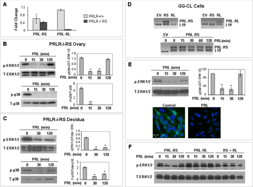
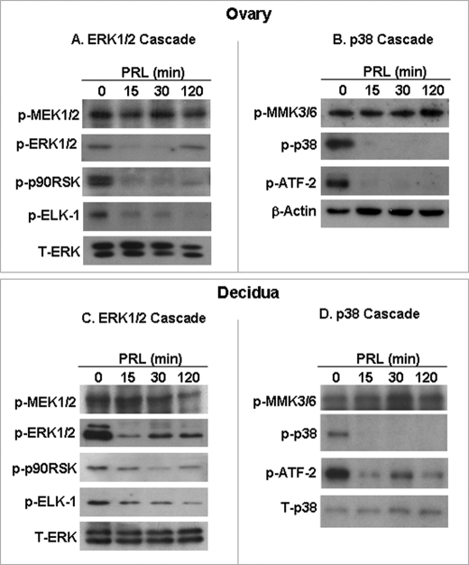
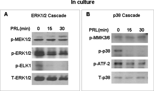
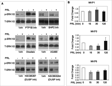
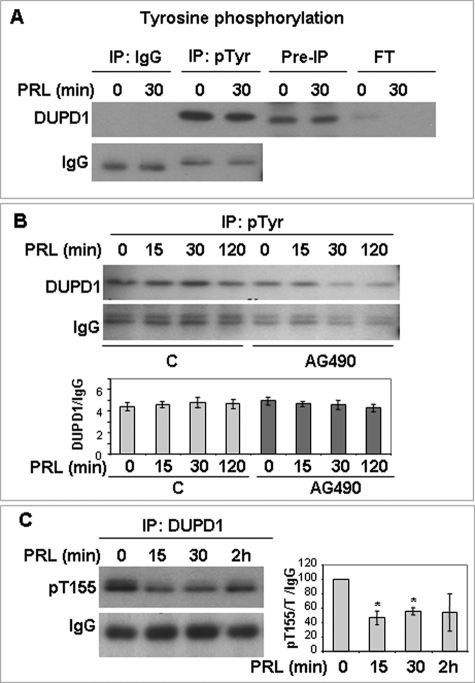
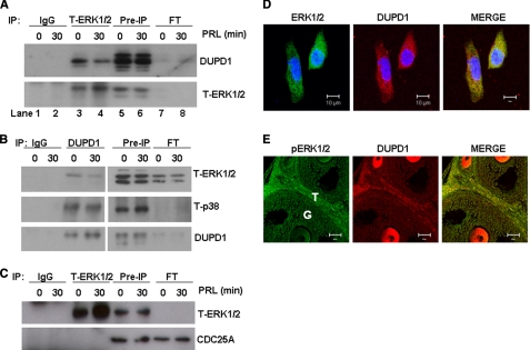
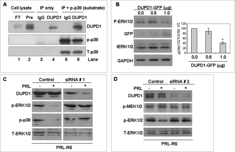
Prolactin (PRL) is essential for normal reproduction and signals through two types of receptors, the short (PRL-RS) and long (PRL-RL) form. We have previously shown that transgenic mice expressing only PRL-RS (PRLR(-/-)RS) display abnormal follicular development and premature ovarian failure. Here, we report that MAPK, essential for normal follicular development, is critically inhibited by PRL in reproductive tissues of PRLR(-/-)RS mice. Consequently, the phosphorylation of MAPK downstream targets are also markedly inhibited by PRL without affecting immediate upstream kinases, suggesting involvement of MAPK specific phosphatase(s) in this inhibition. Similar results are obtained in a PRL-responsive ovary-derived cell line (GG-CL) that expresses only PRL-RS. However, we found the expression/activation of several known MAPK phosphatases not to be affected by PRL, suggesting a role of unidentified phosphatase(s). We detected a 27-kDa protein that binds to the intracellular domain of PRL-RS and identified it as dual specific phosphatase DUPD1. PRL does not induce expression of DUDP1 but represses its phosphorylation on Thr-155. We also show a physical association of this phosphatase with ERK1/2 and p38 MAPK. Using an in vitro phosphatase assay and overexpression studies, we established that DUPD1 is a MAPK phosphatase. Dual specific phosphatase inhibitors as well as siRNA to DUPD1, completely prevent PRL-mediated MAPK inhibition in ovarian cells. Our results strongly suggest that deactivation of MAPK by PRL/PRL-RS contributes to the severe ovarian defect in PRLR(-/-)RS mice and demonstrate the novel association of PRL-RS with DUPD1 and a role for this phosphatase in MAPK deactivation.


Figures

FIGURE 1.
FIGURE 1.
PRL mediated inhibition of MAPK in ovary, decidua, and GG-CL cells expressing only PRL-RS. A, PRL-RS and PRL-RL expression were examined by qPCR in ovaries of wild type (PRLR+/+) and PRLR−/−RS mice. The expression of PRL-RS was not significantly different from wild type, whereas expression of PRL-RL was not detected in PRLR−/−RS ovaries. B and C, pseudopregnant PRLR−/−RS mice were injected with ergocryptine (200 μg, subcutaneously) to inhibit endogenous PRL secretion and were treated 6 h later with a single IP injection of PRL (60 μg). Activation status of ERK1/2 and p38 MAPK were measured by Western blot analysis using phosphospecific ERK1/2 (Thr-202/Tyr-204) and p38 MAPK (Thr-180/Tyr-182) antibodies in either ovaries or decidua. Total ERK1/2 and total p38 were used as loading controls. D, GG-CL cells were transfected with either PRL-RS or PRL-RL expression vectors, and specific expression of either receptor was analyzed by RT-PCR (upper panel). PRL treatment had no effect on PRL-RS expression (lower panel). E, activation status of ERK1/2 was measured by Western blot analysis using phospho-antibodies against ERK1/2 in GG-CL cells. Total ERK1/2 was used as loading control. Density of p-ERK was plotted against T-ERK (upper panel). GG-CL cells transfected with PRL-RS were treated with either PRL (1 μg/ml) or vehicle, and localization of active ERK1/2 was analyzed by immunocytochemistry as described under “Experimental Procedures.” Lower panel, green, p-ERK1/2; blue, DAPI. F, GG-CL cells transfected with PRL-RS, PRL-RL, or both were treated with PRL for different time points as indicated. Activation status of ERK1/2 was measured by Western blot analysis using phosphospecific ERK1/2 (Thr-202/Tyr-204) antibody. The values are expressed as the means ± S.E. (n = 3). *, p < 0.05; **, p < 0.01 versus 0 min.
FIGURE 2.
FIGURE 2.
Downstream targets of MAPK are affected by PRL/PRL-RS but not the upstream kinases in PRLR−/−RS mice. Decidualization in pseudopregnant PRLR−/−RS mice was induced as described under “Experimental Procedures.” PRLR−/−RS mice were injected with ergocryptine (200 μg, subcutaneously) for 6 h followed by a single IP injection of PRL (60 μg). ERK1/2 and p38 MAPK cascades were examined by Western blot analysis in the ovary (A and B) and decidua (C and D) using phospho-specific antibodies to ELK1, p90RSK, ATF2, MEK1/2, and MKK3/6. Total ERK1/2 or β-actin was used loading control.
FIGURE 3.
FIGURE 3.
Downstream targets of MAPK are affected by PRL/PRL-RS but not the upstream kinases in GG-CL cells expressing PRL-RS. GG-CL cells were transfected with PRL-RS and treated with PRL as indicated. Total protein extracts were analyzed for activated ERK1/2 (A) or activated p38 MAPK pathway (B) by Western analysis using phospho-specific antibodies to ELK1, ATF2, MEK1/2, and MKK3/6. Total ERK1/2 and total p38 were used as loading controls.
FIGURE 4.
FIGURE 4.
Effect of various phosphatase inhibitors on the PRL-mediated inhibition of ERK1/2 and mRNA expression of MKPs. A, GG-CL cells transfected with PRL-RS were treated with PRL (1 μg/ml) for 15 min in the presence of protein-tyrosine phosphatase 1B (PTP1B) inhibitors, SHP2 inhibitor, okadaic acid, AG-490, NSC95397, and NSC663284 as described under “Experimental Procedures.” Activation status of ERK1/2 was measured by Western blot analysis using phospho-antibodies against ERK1/2. Total ERK1/2 was used as loading control. B, GG-CL cells transfected with PRL-RS were treated with PRL (1 μg/ml) for several time points as indicated. The mRNA levels of MKP1, MKP3, and MKP5 were analyzed by qPCR in these cells, and the expression levels were normalized to L19 or 36B4.
FIGURE 5.
FIGURE 5.
Identification of DUPD1 as PRL-RS associated protein. PRL-RS-GST (RS-GST) fusion proteins were prepared as described under “Experimental Procedures.” RS-GST interacting proteins were pulled down using decidual cell extracts from PRLR−/−RS mice treated with PRL (60 μg/animal) or vehicle (A) or GG-CL cells transfected with PRL-RS and treated with PRL (1 μg/ml) for different time points (B). The bands were detected by silver staining. The molecular mass markers (lanes M) were as indicated to determine the size of the bands. As a control, a well of pre-pulldown PRL-RS-GST (RS) was loaded showing a band of 25 kDa. The band (27–30 kDa) pulldown from decidual and GG-CL cell lysates by RS-GST fusion protein is indicated by arrow. C, RS-GST pulldown samples of GG-CL cell lysates were analyzed by Western blot using a polyclonal antibody to DUPD1. To show the specific interaction of DUPD1 with RS-GST, GST alone (C-GST) was used to pull down GG-CL cell lysates as a control. GG-CL cells were transfected with PRL-RS and treated with PRL (1 μg/ml) for different time points. Expression of DUPD1 was measured by qPCR (D) or Western analysis (E). *, p < 0.05 versus 0 min.
FIGURE 6.
FIGURE 6.
DUPD1 is a phosphoprotein. A, GG-CL cells were transfected with PRL-RS and treated with PRL for different time points. Total cell lysates were immunoprecipitated with anti-phosphotyrosine antibody or control IgG and immunoblotted for DUPD1. B, GG-CL cells were transfected with PRL-RS and treated with PRL in the presence or absence of AG490. Total cell lysates were immunoprecipitated with anti-phophotyrosine antibody and immunoblotted for DUPD1. Density of DUPD1 was plotted against IgG. C, GG-CL cells were transfected with PRL-RS and treated with PRL. Total cell lysates were immunoprecipitated with DUPD1 and immunoblotted with an anti-phospho-RXRXX(S/T) antibody. The density of phospho-DUPD1 was plotted against IgG. *, p < 0.05 versus 0 min.
FIGURE 7.
FIGURE 7.
DUPD1 physically associates with ERK1/2 and p38 MAPK. GG-CL cells were transfected with PRL-RS and treated with PRL (1 μg/ml) for 30 min. A, total protein extracts were subjected to IP with T-ERK1/2 or rabbit IgG, and immunocomplexes were resolved on SDS-PAGE, transferred onto PVDF membrane, and immunoblotted against DUPD1 or T-ERK1/2s. Lanes 1 and 2, IP with IgG (0 and 30 min PRL-treated samples); lanes 3 and 4, IP with T-ERK1/2 (0 and 30 min PRL-treated samples); lanes 5 and 6, pre-IP samples (total protein extracts); lanes 7 and 8, flow through after IP (unbound supernatants). B, cells were IP with DUPD1 antibody or goat IgG, and immunocomplexes were resolved and immunoblotted as described for A. C, cells were immunoprecipitated with T-ERK1/2 antibody or goat IgG, and immunocomplexes were resolved and immunoblotted against CDC25A and T-ERK1/2. D, GG-CL cells were transfected with PRL-RS and treated with PRL for 30 min. Co-localization of T-ERK1/2 (green) and DUPD1 (red) was examined by immunocytochemistry using specific antibodies. Blue, DAPI. E, paraffin-embedded ovary sections from PRLR−/−RS mice (2.5 days pregnant) were analyzed for co-localization of active ERK1/2 (green) and DUPD1 (red).
FIGURE 8.
FIGURE 8.
DUPD1 is a MAPK phosphatase. A, total protein lysates of GG-CL cells expressing PRL-RS were subjected to IP with a polyclonal antibody to DUPD1 or control IgG. In vitro phosphatase assay was carried out using these immunoprecipitates and a recombinant active p38 (rp-p38) as substrate. Finally, the proteins were resolved on SDS-PAGE and immunoblotted against p-p38 to examine the remaining substrate. The blot was also reprobed for DUPD1 and T-p38. Lane 1, flow through after the IP; lane 2, pre-IP; lane 3, IP with IgG alone; lane 4, IP with DUPD1 alone; lane 5, IP with IgG and rp-p38 (after the assay); lane 6, IP with DUPD1 and rp-p38 (after the assay). B, GG-CL cells were co-transfected with PRL-RS and increasing doses of DUPD1-GFP expression vectors or empty vectors. Phosphorylation status of ERK1/2 was examined using phospho-specific antibody. Expression of exogenous DUPD1 was measured by Western blot against GFP antibody. Total ERK1/2 and GAPDH were used as loading controls. The density of p-ERK1/2 was plotted against T-ERK1/2. *, p < 0.05 versus 0 μg. C, GG-CL cells were co-transfected with PRL-RS and DUPD1 siRNA (siRNA # 1) or a control siRNA. After 48 h, the cells were treated with or without PRL for 15 min. DUPD1, p-ERK1/2, and p-p38 were measured in the total cell lysates by Western blotting. Total ERK1/2 was used as loading control. D, GG-CL cells were co-transfected with PRL-RS and DUPD1 siRNA (siRNA # 2) or a control siRNA. After 48 h, the cells were treated with or without PRL for 15 min. DUPD1, p-MEK1/2 and p-ERK1/2 were measured in the total cell lysates by Western blotting. Total ERK1/2 was used as a loading control.

Similar articles

Cited by

References

    1. Risk M., Gibori G. (2001) in Prolactin (Horseman N. D. ed) pp. 265–295, Kluwer Academic, Boston, MA
    1. Stocco C., Telleria C., Gibori G. (2007) Endocr. Rev. 28, 117–149 - PubMed
    1. Bachelot A., Binart N. (2007) Reproduction 133, 361–369 - PubMed
    1. Ben-Jonathan N., LaPensee C. R., LaPensee E. W. (2008) Endocr. Rev. 29, 1–41 - PMC - PubMed
    1. Halperin J., Devi S. Y., Elizur S., Stocco C., Shehu A., Rebourcet D., Unterman T. G., Leslie N. D., Le J., Binart N., Gibori G. (2008) Mol. Endocrinol. 22, 513–522 - PMC - PubMed

Publication types

MeSH terms

Substances

LinkOut - more resources