. 2011 Feb 25;286(8):6253-61.
doi: 10.1074/jbc.M110.178210. Epub 2011 Jan 3.

The tumor suppressor RASSF1A prevents dephosphorylation of the mammalian STE20-like kinases MST1 and MST2

Affiliations

The tumor suppressor RASSF1A prevents dephosphorylation of the mammalian STE20-like kinases MST1 and MST2

Cai Guo et al. J Biol Chem. .

Abstract

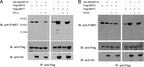
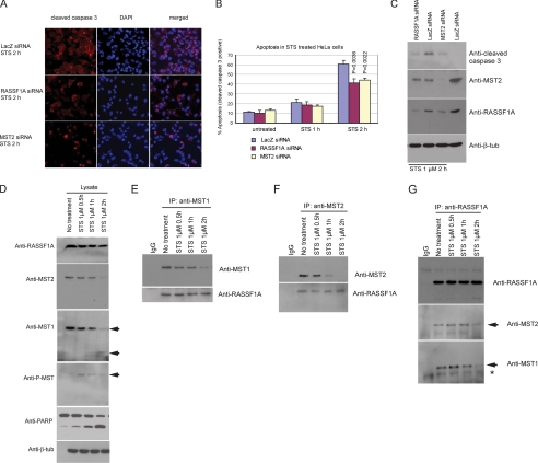
The RASSF1A tumor suppressor protein interacts with the pro-apoptotic mammalian STE20-like kinases MST1 and MST2 and induces their autophosphorylation and activation, but the mechanism of how RASSF1A activates MST1/2 is unclear. Okadaic acid treatment and PP2A knockdown promoted MST1/2 phosphorylation. Data from dephosphorylation assays and reduced activation of MST1/2 seen after RASSF1A depletion suggest that dephosphorylation of MST1/2 on Thr-183 and Thr-180 by PP2A is prevented by RASSF1A, shifting the balance of MST1/2 to the activated autophosphorylated form. In addition to preventing dephosphorylation, RASSF1A also stabilized the MST2 protein. Through binding to MST1/2, RASSF1A supports maintenance of MST1/2 phosphorylation, promoting an active state of the MST kinases and favoring induction of apoptosis. This is one of the first examples of a tumor suppressor acting as an inhibitor of a specific dephosphorylation pathway.


Figures

FIGURE 1.
FIGURE 1.
RASSF1A prevents MST1/2 dephosphorylation. A and B, 5 μg of FLAG-MST1/2 was transfected with 5 μg of HA vector or HA-RASSF1A into 293T cells. Forty-eight hours after transfection, cells were lysed, and the lysates were immunoprecipitated (IP) with EZviewTM Red anti-FLAG resin overnight at 4 °C. In vitro dephosphorylation assays were performed with recombinant PP1 (A) or PP2A (B) added to the anti-FLAG precipitates, followed by the indicated immunoblots (IB).
FIGURE 2.
FIGURE 2.
PP2A induces MST1/2 dephosphorylation counteracted by RASSF1A. A, HeLa cells were either left untreated or were treated with 1 μm okadaic acid (OA) for 1 h. Western blotting was performed using anti-phospho-MST and anti-β-tubulin (anti-β-tub) antibodies. B, HeLa cells were transfected with LacZ control siRNA, PP1 catalytic subunit siRNA, or PP2A α-isoform catalytic subunit siRNA. Seventy-two hours later, cells were treated with 1 μm STS for 1 h to induce apoptosis. After harvesting and lysis, cell lysates were examined by anti-phospho-MST antibody and other blots. The asterisk indicates a nonspecific band. C, phospho-MST levels normalized to total MST protein levels after STS treatment were determined from four independent experiments conducted as shown described for B. Significance was calculated using Student's t test. D, 3.5 μg of FLAG-MST1/2 was transfected with 3.5 μg of HA-PPP2CA, 3.5 μg of HA-RASSF1A, or both. After immunoprecipitation (IP), anti-FLAG precipitates were subject to the indicated immunoblots (IB). Cleaved MST1 is indicated by the asterisk. The two bands in the anti-PPP2CA blot indicate endogenous (lower band) and transfected PPP2CA (upper band).
FIGURE 3.
FIGURE 3.
Effect of RASSF1A depletion on MST1/2 protein levels and phosphorylation and on other phosphoproteins. A, RASSF1A-specific siRNA was used in control and STS-treated HeLa cells to diminish RASSF1A levels, and the effect of the depletion on MST1 and MST2 protein levels and their Thr-183/Thr-180 phosphorylation status was assessed. The asterisk indicates a nonspecific band. B, depletion of RASSF1A leads to a slight reduction of MST2 protein levels in untreated cells. The results were quantitated from three independent experiments, and S.D. is shown. C, depletion of RASSF1A leads to reduction of phospho-MST. A 60% reduction of the RASSF1A protein level (A) is associated with an ∼30% reduction of phospho-MST. The results were quantitated from three independent experiments, and S.D. is shown. D, effect of RASSF1A depletion on levels and phosphorylation status of JNK, p38, ERK1/2, and AKT in untreated and STS-treated HeLa cells. IB, immunoblot; β-tub, β-tubulin.
FIGURE 4.
FIGURE 4.
RASSF1A-MST complexes during apoptosis. A, HeLa cells were transfected with control LacZ siRNA or RASSF1A- or MST2-specific siRNA before treatment with the apoptotic inducer STS. Apoptotic cells were scored at different time points following STS treatment by immunofluorescence staining with antibody against cleaved caspase-3. B, cleaved caspase-3-positive cells were counted from three independent experiments. Between 500 and 2000 cells were counted for each treatment condition and time point, and S.D. is shown. C, Western blot analysis of cells undergoing STS-induced apoptosis after knockdown of RASSF1A or MST2. D, HeLa cells were either left untreated or were treated with 1 μm STS for 0.5, 1, or 2 h. Cells were harvested at different time points, and cell lysates were prepared and subjected to immunoblotting with different antibodies. E, lysates were immunoprecipitated (IP) with anti-MST1 antibody. F, lysates were immunoprecipitated with anti-MST2 antibody. G, lysates were immunoprecipitated with anti-RASSF1A antibody. The immunoprecipitates were fractionated by SDS-PAGE and examined by the indicated blots. The asterisk indicates a nonspecific band. Anti-β-tub, anti-β-tubulin antibody; Anti-PARP, anti-poly(ADP-ribose) polymerase antibody.
FIGURE 5.
FIGURE 5.
Effects of phosphatase knockdown and STS treatment on Hippo pathway components. A, phosphatase knockdown in non-STS-treated cells. HeLa cells were treated with siRNA against PP1 or PP2A catalytic subunits. Lysates were prepared, and Western blotting was performed with the indicated antibodies. The levels of LATS1 and YAP were decreased. B, STS treatment and phosphatase inhibition. HeLa cells transfected with control LacZ siRNA or siRNA against phosphatases were treated with STS for 1 h. Lysates were prepared, and Western blotting was performed with the indicated antibodies. The levels of LATS1 and YAP were decreased. C, YAP in subcellular fractions of HeLa cells after STS treatment. C, cytoplasmic fraction; N, nuclear fraction. Anti-PARP, anti-poly(ADP-ribose) polymerase antibody; Anti-β-tub, anti-β-tubulin antibody.

Similar articles

Cited by

References

    1. Dammann R., Li C., Yoon J. H., Chin P. L., Bates S., Pfeifer G. P. (2000) Nat. Genet. 25, 315–319 - PubMed
    1. Burbee D. G., Forgacs E., Zöchbauer-Müller S., Shivakumar L., Fong K., Gao B., Randle D., Kondo M., Virmani A., Bader S., Sekido Y., Latif F., Milchgrub S., Toyooka S., Gazdar A. F., Lerman M. I., Zabarovsky E., White M., Minna J. D. (2001) J. Natl. Cancer Inst. 93, 691–699 - PMC - PubMed
    1. Dammann R., Schagdarsurengin U., Seidel C., Strunnikova M., Rastetter M., Baier K., Pfeifer G. P. (2005) Histol. Histopathol. 20, 645–663 - PubMed
    1. Agathanggelou A., Cooper W. N., Latif F. (2005) Cancer Res. 65, 3497–3508 - PubMed
    1. Tommasi S., Dammann R., Zhang Z., Wang Y., Liu L., Tsark W. M., Wilczynski S. P., Li J., You M., Pfeifer G. P. (2005) Cancer Res. 65, 92–98 - PubMed

Publication types

MeSH terms