. 2011 Jan 18;108(3):1176-81.
doi: 10.1073/pnas.1013965108. Epub 2011 Jan 3.

Myelination transition zone astrocytes are constitutively phagocytic and have synuclein dependent reactivity in glaucoma

Affiliations

Myelination transition zone astrocytes are constitutively phagocytic and have synuclein dependent reactivity in glaucoma

Judy V Nguyen et al. Proc Natl Acad Sci U S A. .

Abstract

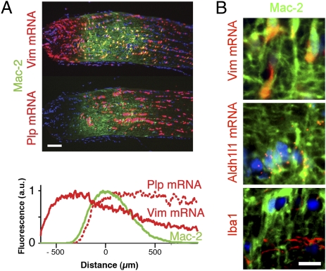
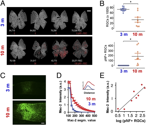
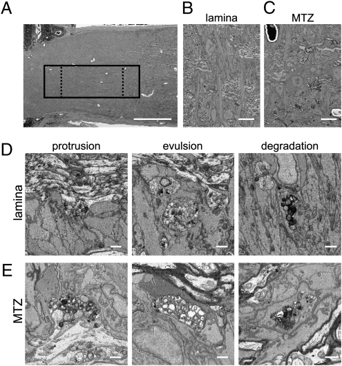
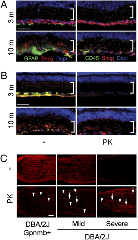
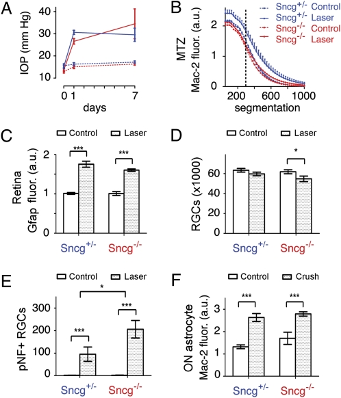
Optic nerve head (ONH) astrocytes have been proposed to play both protective and deleterious roles in glaucoma. We now show that, within the postlaminar ONH myelination transition zone (MTZ), there are astrocytes that normally express Mac-2 (also known as Lgals3 or galectin-3), a gene typically expressed only in phagocytic cells. Surprisingly, even in healthy mice, MTZ and other ONH astrocytes constitutive internalize large axonal evulsions that contain whole organelles. In mouse glaucoma models, MTZ astrocytes further up-regulate Mac-2 expression. During glaucomatous degeneration, there are dystrophic processes in the retina and optic nerve, including the MTZ, which contain protease resistant γ-synuclein. The increased Mac-2 expression by MTZ astrocytes during glaucoma likely depends on this γ-synuclein, as mice lacking γ-synuclein fail to up-regulate Mac-2 at the MTZ after elevation of intraocular pressure. These results suggest the possibility that a newly discovered normal degradative pathway for axons might contribute to glaucomatous neurodegeneration.


Conflict of interest statement

Conflict of interest statement: N.M.-A. and I.S. have a pending patent application on the use of anti-Parkinson disease treatments in glaucoma.

Figures

Fig. 1.
Fig. 1.
A band of Mac-2–expressing astrocytes is located at the postlaminar optic nerve MTZ. (A) Mac-2–expressing cells form a band at the MTZ, as determined by the expression of the astrocyte-specific Vim mRNA and the oligodendrocyte-specific Plp mRNA. Histogram shows the expression of Vim mRNA (solid red), Plp mRNA (dotted red), and Mac-2 (green) as a function of distance from the center of Mac-2 expression. Values represent the mean of mean values from 10 nerves (an average of six sections per nerve). (B) Mac-2–expressing cells at the MTZ are astrocytes, based on the expression of astrocyte-specific Vim and Aldh1l1 mRNAs and lack of expression of the microglial marker Iba1. All images of the ONH in all figures are oriented with the eye to the left of the MTZ. (Scale bars: A, 100 μm; B, 10 μm.)
Fig. 2.
Fig. 2.
Degenerating nerves in DBA/2J mice have a large increase in Mac-2 expression at the MTZ. (A) Retina whole mounts show RGCs, detected by γ-synuclein mRNA (gray), and the position of pNF+ RGCs (red dots). The number of RGCs and pNF+ RGCs are shown below each retina in white and red, respectively. (B) The number of RGCs was lower and the number of pNF+ RGCs higher in the 10-mo relative to the 3-mo DBA/2J mice. *P = 0.003 (Top), *P = 0.01 (Bottom), Mann–Whitney t test. (C) Mac-2 immunoreactivity at the MTZ in representative 3-mo and 10-mo DBA/2J mice. (D) Higher mean intensity of Mac-2 immunoreactivity at the MTZ in 10-mo relative to 3-mo DBA/2J at all segmentation values of Mac-2. For example, at a segmentation of 200, values were approximately sevenfold higher (P < 0.001, two-tailed unpaired t test). Inset: Expression of Mac-2 along the nerve axis, which peaks at the MTZ in nerves of both 3-mo and 10-mo DBA/2J mice. (E) Mac-2 intensity increases linearly for every log increase in the number of pNF+ RGCs (Pearson r = 0.91, P < 0.001). Values represent the mean of nine and eight eyes for the 3-mo and 10-mo mice, respectively. Error bars represent SEM. (Scale bars: A, 500 μm; C, 100 μm.)
Fig. 3.
Fig. 3.
ONH astrocytes internalize large axonal evulsions. (A) Low-power view of the ONH shows the location of one of two volumes acquired (solid box) and the regions used for counting granule accumulations in the lamina and MTZ (dashed boxes). (B) Low-power scanning EM view shows transverse astrocytes at the lamina. (C) Low-power scanning EM view of MTZ shows that astrocyte somata are also transversely oriented at the onset of myelination. In both the lamina (D) and the MTZ (E), granule accumulations exist as swellings and protrusions in axons (Left), as large evulsions completely separated from axons and completely ensheathed by astrocytes (Center), and as accumulations within the cytoplasm of astrocytes (Right). (Scale bars: A, 100 μm; B and C, 10 μm; D and E, 1 μm.)
Fig. 4.
Fig. 4.
Formation of PK-resistant γ-synuclein in glaucomatous retina and ONH. (A) Loss of γ-synuclein immunoreactivity (red) in aged DBA/2J coincides with increases in GFAP in glia and CD45 in microglia (green). Bracket shows the IPL. (B) Immunoreactivity of two γ-synuclein antibodies (red and green) is lost after PK treatment in healthy retinas of 3-mo DBA/2J mice, but much of the immunoreactivity of the C-terminal antibody (red) persists after PK treatment in degenerating retinas of 10-mo DBA/2J mice. (C) Protease-resistant γ-synuclein within the ONH during glaucomatous degeneration. Arrowheads point to examples of PK-resistant γ-synuclein in spheroids, and arrows point to examples of PK-resistant γ-synuclein in elongated processes. (Mac-2 expression in these same ONHs, and the RGCs and pNF+ RGCs in the corresponding retinas, are shown in Fig. S3). (Scale bars: 50 μm.)
Fig. 5.
Fig. 5.
IOP-dependent Mac-2 increase at the MTZ requires γ-synuclein. (A) γ-Synuclein does not affect the amount of IOP elevation produced by translimbal laser photocoagulation. (B) IOP elevation increases Mac-2 expression at the MTZ in mice with one copy of γ-synuclein (Sncg+/−), but does not do so in mice that lack γ-synuclein (Sncg−/−). For example, at the segmentation value of 300, marked by the dotted line, γ-synuclein genotype affects Mac-2 expression (P = 0.01, two-way ANOVA). SEM shown only for Sncg+/− mice for the sake of clarity, although SEM values were similar for Sncg−/− mice. (C) GFAP fluorescence on the retina surface is increased by lasering equally in Sncg+/− and Sncg−/− mice (***P < 0.001, two-way ANOVA). (D) Number of RGCs is decreased by lasering only in Sncg−/− mice (*P < 0.05, two-way ANOVA with Bonferroni posttest). (E) The number of pNF+ RGCs is increased by lasering in both Sncg+/− and Sncg−/− mice, but moreso in Sncg−/− mice. (*P = 0.03, two-way ANOVA effect of genotype; **P < 0.05 and ***P < 0.001, two-way ANOVA with Bonferroni posttest). (F) The Mac-2 fluorescence increase in optic nerve astrocytes (pixels selected by GFAP expression) observed 3 d after crush is not affected by Sncg genotype (***P < 0.001, two-way ANOVA with Bonferroni posttest). Values represent the mean of eight and six eyes for lasered eyes of Sncg+/− and Sncg−/− mice, five and six eyes for nonlasered control eyes in the lasering experiments, and six and seven eyes for Sncg+/− and Sncg−/− mice in optic nerve crush experiments. Error bars represent SEM.

References

    1. Quigley HA, Broman AT. The number of people with glaucoma worldwide in 2010 and 2020. Br J Ophthalmol. 2006;90:262–267. - PMC - PubMed
    1. Inman DM, Horner PJ. Reactive nonproliferative gliosis predominates in a chronic mouse model of glaucoma. Glia. 2007;55:942–953. - PubMed
    1. WoldeMussie E, Ruiz G, Wijono M, Wheeler LA. Neuroprotection of retinal ganglion cells by brimonidine in rats with laser-induced chronic ocular hypertension. Invest Ophthalmol Vis Sci. 2001;42:2849–2855. - PubMed
    1. Son JL, et al. Glaucomatous optic nerve injury involves early astrocyte reactivity and late oligodendrocyte loss. Glia. 2010;58:780–789. - PubMed
    1. Hernandez MR, Miao H, Lukas T. Astrocytes in glaucomatous optic neuropathy. Prog Brain Res. 2008;173:353–373. - PubMed

Publication types