Case Reports
. 2010 Dec 31:16:2955-63.

Congenital hereditary endothelial dystrophy - mutation analysis of SLC4A11 and genotype-phenotype correlation in a North Indian patient cohort

Affiliations
Case Reports

Congenital hereditary endothelial dystrophy - mutation analysis of SLC4A11 and genotype-phenotype correlation in a North Indian patient cohort

Preeti Paliwal et al. Mol Vis. .

Abstract

Purpose: To identify the solute carrier family 4 (sodium borate cotransporter) member 11 (SLC4A11) mutation spectrum and to perform genotype-phenotype correlations in autosomal recessive Congenital Hereditary Endothelial Dystrophy (CHED2) in North Indian patients.

Methods: Twenty-five patients from twenty families clinically diagnosed with autosomal recessive CHED2 were recruited for the study. Clinical parameters such as age at onset, presentation, and pre- and post-operative visual acuities were recorded. Corneal buttons of patients undergoing keratoplasty were analyzed for histopathologic and ultrastructural confirmation. All the affected individuals and 50 unrelated population matched normal controls were screened for underlying sequence changes. Genomic DNA was isolated from peripheral blood samples and all the exons and the 5'-upstream region of the SLC4A11 gene were screened for mutations by direct DNA sequencing.

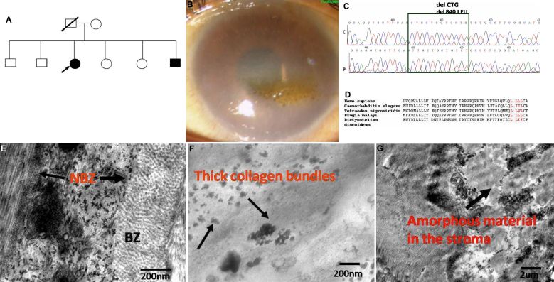
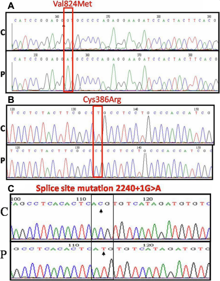
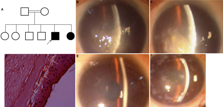
Results: A high degree of consanguinity (9 out of 20 families) was noted. Corneal haze was reported to be present since birth or shortly thereafter in all affected patients. Histology and electron microscopy studies revealed increased thickness of Descemet's membrane, especially of the non-banded zone. Molecular studies revealed one novel homozygous in-frame deletion mutation in two affected siblings from one family and three other previously reported homozygous mutations in 12 patients from 9 families. Mutations were not identified in 11 patients from 11 families. High interfamilial and intrafamilial phenotypic variability was seen among the cohort of patients.

Conclusions: This is the first report on the mutation spectrum and genotype-phenotype correlation in CHED2 patients from North India. The present study detected one novel and three reported changes, adding to the repertoire of mutations in SLC4A11, and recorded a high degree of genetic heterogeneity in CHED2.


Figures

Figure 1
Figure 1
Genotype-phenotype features of the novel SLC4A11 deletion mutation. A: Pedigree of the family showing a novel deletion mutation of one of the four leucine residues c2518-c2520 del CTG in the exon 18 of Solute Carrier Family 4 (sodium borate co-transporter) member 11 (SLC4A11). Filled boxes represent affected individuals. Open boxes represent unaffected individuals. Arrow indicates the proband. B: Slit lamp photomicrographs of the affected individual harboring the novel mutation. The representative clinical photograph shows the presence of marked stromal haze and spheroidal degeneration in the right eye of the proband. C: Partial nucleotide sequence of SLC4A11. The chromatogram of the patient (P) is shown in comparison to control (C). The block marks the four CTG repeats in the control and only three in the patient. The homozygous deletion of CTG residue in the patient can be noted. D: Multiple sequence alignment of SLC4A11 gene from different species. The amino acid leucine (L) at positions 840–843 is conserved over a range of species in the course of evolution, which are highlighted in red. E: Transmission electron micrographs of the affected patient harboring the novel mutation. E: Transmission electron micrograph showing Descemet’s membrane of the CHED2 patient. Descemet’s membrane is thickened, with a normal anterior banded zone and a thickened posterior banded layer (Scale bar 2 μm). F: This panel represents a magnified view of part of the posterior banded layer showing presence of thick collagen bundles indicated by the arrows. G: The disorganized corneal stroma can be noted along with the presence of amorphous material. (Scale bar 2 μm).
Figure 2
Figure 2
Mutations in SLC4A11 causing CHED2. A: Partial nucleotide sequence SLC4A11. The chromatograms of the patients (P) are shown in comparison to controls (C). The homozygous G >A substitution is marked by the block. The block denotes the nucleotide with a missense mutation resulting in amino acid substitution of valine at amino acid position 824 with methionine. B: The homozygous T>C substitution is marked by the block. The block denotes the nucleotide with a missense mutation resulting in amino acid substitution of cysteine at amino acid position 386 with arginine. C: The homozygous G>A substitution is marked by the block. The block denotes the nucleotide with a missense mutation resulting in a splice site mutation c.2240+1G>A.
Figure 3
Figure 3
Family Q1 showing variable phenotype. A: Pedigree of the family showing the splice site mutation c.2240+1G>A and variable phenotypic presentation of the affected members. Filled boxes represent affected individuals. Open boxes represent unaffected individuals. Arrow indicates the proband. A double line indicates presence of consanguinity in the family. B, C: Representative slit lamp photomicrographs of the proband with a homozygous splice site mutation c.2240+1G>A. The representative clinical photographs of right (B) and left eye (C) of the proband shows the presence of the typical ground glass appearance of the cornea seen in autosomal recessive CHED. D: shows the presence of apple green birefringence on staining with Congo-red and viewing under polarized filter, marked by arrows. E: The slit lamp photomicrograph of the right eye of the affected sibling had marked stromal haze. F: The clinical photomicrograph of the mother shows the endothelial deposits (marked by arrows) with stromal haze. A few epithelial deposits are also seen.

References

    1. Ehlers N, Módis L, Møller-Pedersen T. A morphological and functional study of congenital hereditary endothelial dystrophy. Acta Ophthalmol Scand. 1998;76:314–8. - PubMed
    1. Weiss JS, Møller HU, Lisch W, Kinoshita S, Aldave AJ, Belin MW, Kivelä T, Busin M, Munier FL, Seitz B, Sutphin J, Bredrup C, Mannis MJ, Rapuano CJ, Van Rij G, Kim EK, Klintworth GK. The IC3D classification of the corneal dystrophies. Cornea. 2008;27:S1–83. - PMC - PubMed
    1. Toma NM, Ebenezer ND, Inglehearn CF, Plant C, Ficker LA, Bhattacharya SS. Linkage of congenital hereditary endothelial dystrophy to chromosome 20. Hum Mol Genet. 1995;4:2395–8. - PubMed
    1. Hand CK, Harmon DL, Kennedy SM, FitzSimon JS, Collum LMT, Parfrey NA. Localization of the gene for autosomal recessive congenital hereditary endothelial dystrophy (CHED2) to chromosome 20 by homozygosity mapping. Genomics. 1999;61:1–4. - PubMed
    1. Vithana EN, Morgan P, Sundaresan P, Ebenezer ND, Tan DT, Mohamed MD, Anand S, Khine KO, Venkataraman D, Yong VH, Salto-Tellez M, Venkatraman A, Guo K, Hemadevi B, Srinivasan M, Prajna V, Khine M, Casey JR, Inglehearn CF, Aung T. Mutations in sodium-borate co-transporter SLC4A11 cause recessive congenital hereditary endothelial dystrophy, CHED2. Nat Genet. 2006;38:755–7. - PubMed

Publication types

MeSH terms

Supplementary concepts

LinkOut - more resources