. 2010 Dec;3(12):22-30.

Electro-optical Synergy Technique: A New and Effective Nonablative Approach to Skin Aging

Electro-optical Synergy Technique: A New and Effective Nonablative Approach to Skin Aging

Moetaz El-Domyati et al. J Clin Aesthet Dermatol. 2010 Dec.

Abstract

Objectives: Electro-optical synergy technology is one of the most recently described methods for nonablative skin rejuvenation. The aim of this study is to evaluate the effects of electro-optical synergy on connective tissue composition by histological and immunohistochemical techniques coupled with computerized morphometric analysis.

Design: A prospective clinical study.

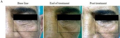
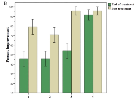
Participants: Six volunteers with Fitzpatrick skin types 3 to 4 and Glogau class I to II wrinkles were subjected to three months (6 sessions at 2-week intervals) of electro-optical synergy treatment.

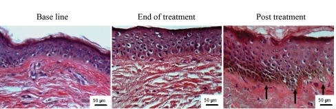
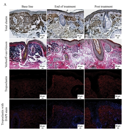
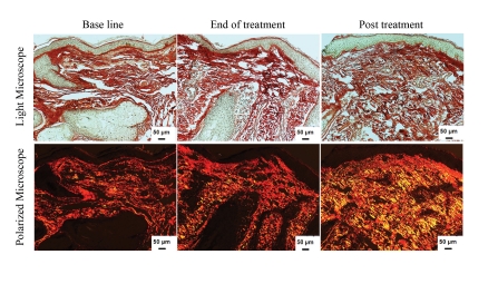
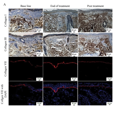
Measurements: Standard photographs and skin biopsies were obtained at baseline as well as three and six months after the start of treatment. The authors performed quantitative evaluation of total elastin, tropoelastin, collagen types I, III, and VII, and newly synthesized collagen.

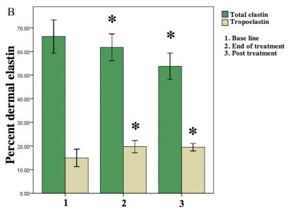
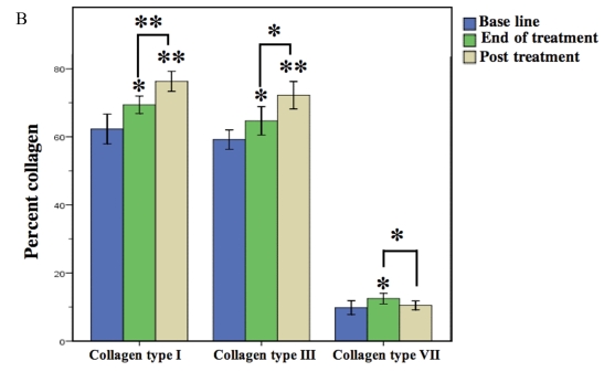
Results: Noticeable clinical and histological improvement was observed after electro-optical synergy treatment. A statistically significant increase in the means of collagen types I, III, and VII, as well as newly synthesized collagen, together with increased levels of tropoelastin, were detected, while the mean level of total elastin was significantly decreased at the end of treatment and three months post-treatment.

Conclusion: Electro-optical synergy is an effective treatment for contouring facial skin laxity. This modality stimulates the repair processes and reverses the clinical, as well as the histopathological, signs of aging with the advantage of being a relatively risk-free procedure with minimal patient recovery time.


Figures

Figures 1A and 1B
Figures 1A and 1B
Clinical appearance of volunteers treated with ELOS. (A) Representative photographs of the periorbital area at baseline, end of treatment, and three months post-treatment. (B) The volunteers' evaluation rates showed the mean percent improvement of skin tightening (column 1), skin texture (column 2), wrinkles (column 3), and overall satisfaction (column 4) at end of treatment and three months post-treatment as compared to baseline. The values are mean ± standard deviation.
Figures 1A and 1B
Figures 1A and 1B
Clinical appearance of volunteers treated with ELOS. (A) Representative photographs of the periorbital area at baseline, end of treatment, and three months post-treatment. (B) The volunteers' evaluation rates showed the mean percent improvement of skin tightening (column 1), skin texture (column 2), wrinkles (column 3), and overall satisfaction (column 4) at end of treatment and three months post-treatment as compared to baseline. The values are mean ± standard deviation.
Figure 2
Figure 2
Enhanced epidermal thickness in response to ELOS treatment. Skin biopsies from a volunteer before and after ELOS treatment. Tissue sections, stained with H&E, show epidermal hyperplasia with increased thickness of the granular cell layer associated with the development of rete ridges (arrows).
Figures 3A and 3B
Figures 3A and 3B
Decrease in dermal elastin upon ELOS treatment. (A) Skin tissues at baseline, at the end, and three months post-ELOS treatment were immunostained for total elastin (top row) and stained with Verhoeff van Gieson (2nd row), showing a decrease in elastic tissue. An area, as demarcated by a rectangle, was used to assess elastin level (see B). *=Refers to grenz zone. Immunofluorescence staining of tropoelastin (3rd row) shows increased deposition of newly synthesized tropoelastin in dermis. The same tissues were counter stained blue for nuclei with DAPI (4th row). (B) Percent of dermis occupied by elastin and tropoelastin showing significant changes after treatment. The values are mean ± standard deviation (* = p<0.05, in comparison to the baseline).
Figures 3A and 3B
Figures 3A and 3B
Decrease in dermal elastin upon ELOS treatment. (A) Skin tissues at baseline, at the end, and three months post-ELOS treatment were immunostained for total elastin (top row) and stained with Verhoeff van Gieson (2nd row), showing a decrease in elastic tissue. An area, as demarcated by a rectangle, was used to assess elastin level (see B). *=Refers to grenz zone. Immunofluorescence staining of tropoelastin (3rd row) shows increased deposition of newly synthesized tropoelastin in dermis. The same tissues were counter stained blue for nuclei with DAPI (4th row). (B) Percent of dermis occupied by elastin and tropoelastin showing significant changes after treatment. The values are mean ± standard deviation (* = p<0.05, in comparison to the baseline).
Figure 4
Figure 4
Increase in newly synthesized collagen content in response to ELOS treatment. Representative examples of skin biopsies stained with picrosirius red viewed under bright field microscope (top panels) and polarized field (bottom panels). Bright field captures total collagen content. Polarized light shows a yellow-to-orange birefringence with newly synthesized collagen in yellow and total collagen in red. Note the increase in newly synthesized collagen as reflected by the yellow color after ELOS treatment.
Figures 5A and 5B
Figures 5A and 5B
Increase in collagen content in response to ELOS. (A) Immunohistochemical staining of skin tissues at baseline, at the end, and post-ELOS treatment for collagen type I (1st row) and collagen type III (2nd row). An increase in collagen type VII expression (red; 3rd row) was observed after ELOS treatments compared to baseline, and then decreased three months post-treatment. Nuclei stained in blue with DAPI (4th row). (B) Collagen level was measured and values presented as a percentage of dermis occupied by collagen. The values are mean ± standard deviation (* = p<0.05;** = p<0.001).
Figures 5A and 5B
Figures 5A and 5B
Increase in collagen content in response to ELOS. (A) Immunohistochemical staining of skin tissues at baseline, at the end, and post-ELOS treatment for collagen type I (1st row) and collagen type III (2nd row). An increase in collagen type VII expression (red; 3rd row) was observed after ELOS treatments compared to baseline, and then decreased three months post-treatment. Nuclei stained in blue with DAPI (4th row). (B) Collagen level was measured and values presented as a percentage of dermis occupied by collagen. The values are mean ± standard deviation (* = p<0.05;** = p<0.001).

Similar articles

Cited by

References

    1. Kligman LH, Kligman AM. The nature of photoaging: its prevention and repair. Photodermatol. 1986;3:215–227. - PubMed
    1. El-Domyati M, Attia S, Saleh F, et al. Intrinsic aging vs. photoaging: a comparative histopathological, immuno-histochemical, and ultrastructural study of skin. Exp Dermatol. 2002;11:398–405. - PubMed
    1. Helfrich YR, Sachs DL, Voorhees JJ. Overview of skin aging and photoaging. Dermatol Nurs. 2008;20:177–183. - PubMed
    1. Uitto J. The role of elastin and collagen in cutaneous aging: intrinsic aging versus photoexposure. J Drugs Dermatol. 2008;7:S12–S16. - PubMed
    1. Sadick NS, Weiss R. Intense pulsed-light photorejuvenation. Semin Cutan Med Surg. 2002;21:280–287. - PubMed

LinkOut - more resources