Molecular basis of differential B-pentamer stability of Shiga toxins 1 and 2
- PMID: 21203383
- PMCID: PMC3010993
- DOI: 10.1371/journal.pone.0015153
Molecular basis of differential B-pentamer stability of Shiga toxins 1 and 2
Abstract
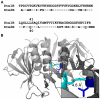
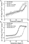
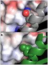
Escherichia coli strain O157:H7 is a major cause of food poisoning that can result in severe diarrhea and, in some cases, renal failure. The pathogenesis of E. coli O157:H7 is in large part due to the production of Shiga toxin (Stx), an AB(5) toxin that consists of a ribosomal RNA-cleaving A-subunit surrounded by a pentamer of receptor-binding B subunits. There are two major isoforms, Stx1 and Stx2, which differ dramatically in potency despite having 57% sequence identity. Animal studies and epidemiological studies show Stx2 is associated with more severe disease. Although the molecular basis of this difference is unknown, data suggest it is associated with the B-subunit. Mass spectrometry studies have suggested differential B-pentamer stability between Stx1 and Stx2. We have examined the relative stability of the B-pentamers in solution. Analytical ultracentrifugation using purified B-subunits demonstrates that Stx2B, the more deadly isoform, shows decreased pentamer stability compared to Stx1B (EC(50) = 2.3 µM vs. EC(50) = 0.043 µM for Stx1B). X-ray crystal structures of Stx1B and Stx2B identified a glutamine in Stx2 (versus leucine in Stx1) within the otherwise strongly hydrophobic interface between B-subunits. Interchanging these residues switches the stability phenotype of the B-pentamers of Stx1 and Stx2, as demonstrated by analytical ultracentrifugation and circular dichroism. These studies demonstrate a profound difference in stability of the B-pentamers in Stx1 and Stx2, illustrate the mechanistic basis for this differential stability, and provide novel reagents to test the basis for differential pathogenicity of these toxins.
Conflict of interest statement
Figures


Similar articles
-
Stability of the homopentameric B subunits of shiga toxins 1 and 2 in solution and the gas phase as revealed by nanoelectrospray fourier transform ion cyclotron resonance mass spectrometry.J Am Soc Mass Spectrom. 2005 Dec;16(12):1957-68. doi: 10.1016/j.jasms.2005.07.016. Epub 2005 Oct 20. J Am Soc Mass Spectrom. 2005. PMID: 16242954
-
Baicalein Inhibits Stx1 and 2 of EHE: Effects of Baicalein on the Cytotoxicity, Production, and Secretion of Shiga Toxins of Enterohaemorrhagic Escherichia coli.Toxins (Basel). 2019 Aug 29;11(9):505. doi: 10.3390/toxins11090505. Toxins (Basel). 2019. PMID: 31470657 Free PMC article.
-
Structure of shiga toxin type 2 (Stx2) from Escherichia coli O157:H7.J Biol Chem. 2004 Jun 25;279(26):27511-7. doi: 10.1074/jbc.M401939200. Epub 2004 Apr 9. J Biol Chem. 2004. PMID: 15075327
-
A Conserved Structural Motif Mediates Retrograde Trafficking of Shiga Toxin Types 1 and 2.Traffic. 2015 Dec;16(12):1270-87. doi: 10.1111/tra.12338. Epub 2015 Nov 2. Traffic. 2015. PMID: 26420131 Free PMC article.
-
Do the A subunits contribute to the differences in the toxicity of Shiga toxin 1 and Shiga toxin 2?Toxins (Basel). 2015 Apr 29;7(5):1467-85. doi: 10.3390/toxins7051467. Toxins (Basel). 2015. PMID: 25938272 Free PMC article. Review.
Cited by
-
Cryo-EM structures of engineered Shiga toxin-based immunogens capable of eliciting neutralizing antibodies with therapeutic potential against hemolytic uremic syndrome.Protein Sci. 2025 Jun;34(6):e70178. doi: 10.1002/pro.70178. Protein Sci. 2025. PMID: 40411437
-
Cytotoxic Effects of Recombinant StxA2-His in the Absence of Its Corresponding B-Subunit.Toxins (Basel). 2021 Apr 26;13(5):307. doi: 10.3390/toxins13050307. Toxins (Basel). 2021. PMID: 33925951 Free PMC article.
-
Shiga Toxins as Multi-Functional Proteins: Induction of Host Cellular Stress Responses, Role in Pathogenesis and Therapeutic Applications.Toxins (Basel). 2016 Mar 17;8(3):77. doi: 10.3390/toxins8030077. Toxins (Basel). 2016. PMID: 26999205 Free PMC article. Review.
-
Excess A-subunits of Shiga toxin 2a are produced in enterohemorrhagic Escherichia coli.Sci Rep. 2025 May 14;15(1):16712. doi: 10.1038/s41598-025-01342-2. Sci Rep. 2025. PMID: 40368985 Free PMC article.
-
Shiga toxin 1 is more dependent on the P proteins of the ribosomal stalk for depurination activity than Shiga toxin 2.Int J Biochem Cell Biol. 2011 Dec;43(12):1792-801. doi: 10.1016/j.biocel.2011.08.018. Epub 2011 Sep 3. Int J Biochem Cell Biol. 2011. PMID: 21907821 Free PMC article.
References
-
- Centers for Disease Control and Prevention. Disease listing, enterohemorrhagic E. coli. 2005.
-
- Mead PS, Griffin PM. Escherichia coli O157:H7. Lancet. 1998;352(9135):1207–1212. - PubMed
-
- Boyce TG, Swerdlow DL, Griffin PM. Escherichia coli O157:H7 and the hemolytic-uremic syndrome. N Engl J Med. 1995;333(6):364–368. - PubMed
-
- Siegler RL. The hemolytic uremic syndrome. Pediatr Clin North Am. 1995;42(6):1505–1529. - PubMed
Publication types
MeSH terms
Substances
Grants and funding
LinkOut - more resources
Full Text Sources
Other Literature Sources

