. 2010 Dec 23;5(12):e15736.
doi: 10.1371/journal.pone.0015736.

In vivo imaging of HIF-active tumors by an oxygen-dependent degradation protein probe with an interchangeable labeling system

Affiliations

In vivo imaging of HIF-active tumors by an oxygen-dependent degradation protein probe with an interchangeable labeling system

Takahiro Kuchimaru et al. PLoS One. .

Abstract

Hypoxia-inducible factor (HIF) functions as a master transcriptional regulator for adaptation to hypoxia by inducing adaptive changes in gene expression for regulation of proliferation, angiogenesis, apoptosis and energy metabolism. Cancers with high expression of the alpha subunit of HIF (HIFα) are often malignant and treatment-resistant. Therefore, the development of a molecular probe that can detect HIF activity has great potential value for monitoring tumor hypoxia. HIF prolyl hydroxylases (HPHDs) act as oxygen sensors that regulate the fate of HIFα protein through its oxygen-dependent degradation (ODD) domain. We constructed a recombinant protein PTD-ODD-HaloTag (POH) that is under the same ODD regulation as HIFα and contains protein transduction domain (PTD) and an interchangeable labeling system. Administration of near-infrared fluorescently labeled POH (POH-N) to mouse models of cancers allowed successful monitoring of HIF-active regions. Immunohistochemical analysis for intratumoral localization of POH probe revealed its specificity to HIF-active cells. Furthermore, lack of the PTD domain or a point mutation in the ODD domain abrogated the specificity of POH-N to HIF-active cells. Overall results indicate that POH is a practical probe specific to HIF-active cell in cancers and suggest its large potential for imaging and targeting of HIF-related diseases.


Conflict of interest statement

Competing Interests: The authors have declared that no competing interests exist.

Figures

Figure 1
Figure 1. Preparation of POH-N probes.
(A) Structure of the POH-N probes. POH fusion protein is composed of PTD, ODD and HaloTag domains. HaloTag ligand labeled with NIRF dye (HL-N) is covalently bound to POH via HaloTag domain to form POH-N probe. (B) SDS-PAGE of prepared probes. PTD-ODD-HaloTag (POH), POmH (POH with a point mutation in the ODD domain) and ODD-HaloTag (OH) fusion proteins were mixed with Alexa Fluor 750- or IR800-labeled HaloTag ligand (HL-A or HL-I) and then unbound HL-A or HL-I was removed by column purification. Probes resolved by SDS-PAGE were stained with Coomassie Blue (upper panel). Fluorescence was scanned by IVIS using excitation (Ex) and emission (Em) filters indicated below the gels (bottom panel). (C) Mechanism of selective imaging for HIF (+) cells. In cells without HIFα (HIF (–) cells), POH-N is immediately degraded through HPHD-mediated ODD and the resultant POH-N fragments and HL-N are diffused from the cells. In cells with HIFα [HIF (+) cells], POH-N is more stable and a clear contrast between cells with versus without HIF activity can be achieved. (D) POH-A and POH-I were added to the culture medium of SUIT2/HRE-Luc cells. After incubation for 1 h under normoxic conditions, the cells were washed and fluorescence intensity was measured (uptake). The cells were further cultured under normoxic conditions for 30 min and fluorescence intensity was measured again (residual). *P<0.01.
Figure 2
Figure 2. In vivo optical imaging of HIF-1 activity in subcutaneous cancers with POH probe and its PTD function in vitro and in vivo.
(A) Membrane permeability of POH-I and OH-I in SUIT-2/HRE-Luc cells. The cells cultured under normoxic or hypoxic conditions were co-cultured with POH-I or OH-I for 1 h and then the fluorescent intensity of cell lysates was measured. The experiments were repeated three times. (B) Representative in vivo image after 2 nmol of POH-I or OH-I administration. Nude mice carrying SUIT-2/HRE-Luc xenografts in both forefeet were imaged at the indicated times after probe injection. The right panel (HIF-1) shows bioluminescence images at 24 h. The bottom panels were magnified images of tumor area of middle panels. (C) The relative fluorescence intensity of the tumor to muscle (T/B ratio). Fluorescence intensities of the SUIT-2/HRE-Luc xenografts and the muscle of the hind foot were measured at the indicated times after POH-I administration. *P<0.01, n = 5.
Figure 3
Figure 3. Characterization of POH probe in vitro and in vivo.
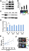
(A and B) Correlation of POH stability and the expression of HIF-1α. The SUIT-2/HRE-Luc cells (1.5×10⁵) cultured under normoxic (N) or hypoxic (H) conditions with 250 µM CoCl2 (Co), a HPHD inhibitor, were treated with POH protein and analyzed by western blotting (representative blot is shown) (A) and measurement of fluorescent intensity (B) with an Ex/Em wavelength of 710 nm/800 nm (P<0.05, n = 3). Representative fluorescence images are shown below the graphs. (C and D) PHD2 regulation of POH stability. The three independent SUIT-2/HRE-Luc/PHD2-KD cell lines (KD1, KD2 and KD3) with reduced PHD2 protein expression (C), which were isolated after stable transfection of a PHD2-specific shRNA plasmid into SUIT-2/HRE-Luc cells, were cultured under normoxic (N) or hypoxic (H) conditions and were treated with POH-I for 1 h. The cells were cultured in fresh medium and the probes remained in the cells was estimated by the measurement of fluorescent intensity of cell lysates. The experiments were repeated three times and a relative value (hypoxia  = 1) means ± SEM are shown (D). *P<0.05 (vs. normoxic condition). (E) Correlation between tumor bioluminescence intensity and the T/B ratio (relative fluorescence intensity of the tumors to the muscle). Bioluminescence and fluorescent intensities of xenografts were measured 24 h after POH-I administration. Bioluminescence intensity and T/B ratio show the coefficient value of 0.7767. n = 29. (F) Representative ex vivo bioluminescence (HIF-1) and fluorescence (POH-I) images of excised subcutaneous tumors. Nude mice carrying SUIT-2/HRE-Luc xenografts in both forefeet were injected with POH-I and dissected 24 h after probe administration. Bar  = 10 mm.
Figure 4
Figure 4. ODD function of POH probe in vitro and in vivo.
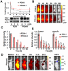
(A) Time-dependent degradation of POH-I and POmH-I in HIF (-) cells. SUIT-2/HRE-Luc cells cultured under normoxic conditions were co-cultured with POH-I or POmH-I for 1 h. The cells were cultured in fresh medium and the probes remained in the cells was estimated by the measurement of fluorescent intensity of cell lysates at the represented time-points, shown by the relative value (0 h = 1). Degradation of POH and POmH protein was also detected by western blotting (lower panels). (B and C) Biodistribution of POH-I and POmH-I in the whole body. Mice without xenografting were imaged at the indicated time after 2 nmol intravenous administration of POH-I or POmH-I (B). Fluorescent intensity of the whole mouse was measured at the indicated time after administration of POH-I or POmH-I and relative fluorescent intensities are shown (C). *P<0.01 (n = 4). (D) In vivo imaging with POmH-I. Nude mice carrying SUIT-2/HRE-Luc xenografts in both forefeet were injected with 2 nmol of POmH-I and images were taken at the indicated times. The right panel (HIF-1) shows bioluminescence images at 24 h. (E) Fluorescence intensities of tumors and the muscle after POH-I (n = 4) or POmH-I (n = 6) administration were measured at the indicated time and relative fluorescent intensities are shown. *P<0.01.
Figure 5
Figure 5. Intratumoral localization of POH probe.
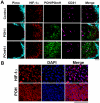
(A) Immunohistochemical analyses of SUIT-2/HRE-Luc xenografts. Tumors were removed 6 h after POH-I or POmH-I administration and examined for sever hypoxic regions (Pimo), HIF (+) cells (HIF-1α), probes (POH/POmH) and blood vessel (CD31). Bars  = 100 µm. (B) Cellular localization of HIF-1α and POH. Tumors were excised 6 h after probe injection and examined for cell nuclei (DAPI) and HIF-1α or POH. Bars  = 100 µm.
Figure 6
Figure 6. In vivo optical imaging of HIF-1 activity in an orthotopic pancreatic cancer model with POH probes.
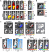
(A) In vivo imaging of pancreatic cancer. Nude mice were orthotopically injected with SUIT-2/HRE-Luc cells and 11 days (upper panels) or 19 days (bottom panels) later the mice were intraperitoneally injected with POH-I or POH-A and imaged at the indicated times after probe administration. Bioluminescence images (HIF-1) of the mice 24 h after probe administration are shown in the right panels. (B and C) Ex vivo imaging of pancreatic cancer. The mice shown in the upper and lower panels of (A) were analyzed by ex vivo imaging and shown in (B) and (C), respectively. Fluorescence (POH-I or POH-A) and bioluminescence (HIF-1) images at 24 h are shown. Bar  = 10 mm (B) or 5 mm (C). (D) Three dimensional analysis of acquired fluorescent (POH-A) and bioluminescent (HIF-1) images. A mouse with pancreatic cancer was injected with POH-A 10 days after orthotopic transplantation and were imaged 24 h after POH-A injection. (E) A mouse with pancreatic cancer was injected with POH-I 11 and 19 days after orthotopic transplantation. Fluorescence (POH-I) and bioluminescence (HIF-1) images were taken 24 h after POH-I injection on the indicated days.

Similar articles

Cited by

References

    1. Semenza GL. Targeting HIF-1 for cancer therapy. Nat Rev Cancer. 2003;3:721–732. - PubMed
    1. Brown JM, Wilson WR. Exploiting tumour hypoxia in cancer treatment. Nat Rev Cancer. 2004;4:437–447. - PubMed
    1. Semenza GL. Hypoxia-inducible factor 1 and cancer pathogenesis. IUBMB Life. 2008;60:591–597. - PubMed
    1. Kaelin WG. Proline hydroxylation and gene expression. Annu Rev Biochem. 2005;74:115–128. - PubMed
    1. Schofield CJ, Ratcliffe PJ. Oxygen sensing by HIF hydroxylases. Nat Rev Mol Cell Biol. 2004;5:343–354. - PubMed

Publication types