. 2010 Dec 23;5(12):e15782.
doi: 10.1371/journal.pone.0015782.

Structure-function relationship of cytoplasmic and nuclear IκB proteins: an in silico analysis

Affiliations

Structure-function relationship of cytoplasmic and nuclear IκB proteins: an in silico analysis

Balachandran Manavalan et al. PLoS One. .

Abstract

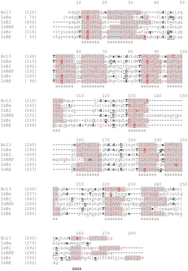
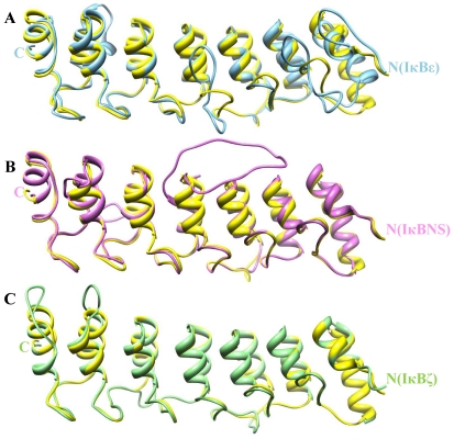
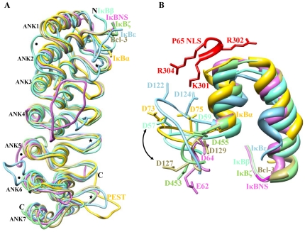
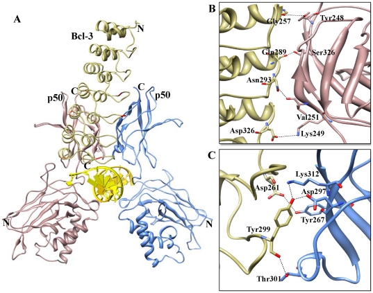
Cytoplasmic IκB proteins are primary regulators that interact with NF-κB subunits in the cytoplasm of unstimulated cells. Upon stimulation, these IκB proteins are rapidly degraded, thus allowing NF-κB to translocate into the nucleus and activate the transcription of genes encoding various immune mediators. Subsequent to translocation, nuclear IκB proteins play an important role in the regulation of NF-κB transcriptional activity by acting either as activators or inhibitors. To date, molecular basis for the binding of IκBα, IκBβ and IκBζ along with their partners is known; however, the activation and inhibition mechanism of the remaining IκB (IκBNS, IκBε and Bcl-3) proteins remains elusive. Moreover, even though IκB proteins are structurally similar, it is difficult to determine the exact specificities of IκB proteins towards their respective binding partners. The three-dimensional structures of IκBNS, IκBζ and IκBε were modeled. Subsequently, we used an explicit solvent method to perform detailed molecular dynamic simulations of these proteins along with their known crystal structures (IκBα, IκBβ and Bcl-3) in order to investigate the flexibility of the ankyrin repeat domains (ARDs). Furthermore, the refined models of IκBNS, IκBε and Bcl-3 were used for multiple protein-protein docking studies for the identification of IκBNS-p50/p50, IκBε-p50/p65 and Bcl-3-p50/p50 complexes in order to study the structural basis of their activation and inhibition. The docking experiments revealed that IκBε masked the nuclear localization signal (NLS) of the p50/p65 subunits, thereby preventing its translocation into the nucleus. For the Bcl-3- and IκBNS-p50/p50 complexes, the results show that Bcl-3 mediated transcription through its transactivation domain (TAD) while IκBNS inhibited transcription due to its lack of a TAD, which is consistent with biochemical studies. Additionally, the numbers of identified flexible residues were equal in number among all IκB proteins, although they were not conserved. This could be the primary reason for their binding partner specificities.


Conflict of interest statement

Competing Interests: The authors have declared that no competing interests exist.

Figures

Figure 1
Figure 1. Structure-based sequence alignments of ARD domains.
The JOY program was used to annotate the alignments for Bcl-3, IκBα, IκBζ, IκBNS, IκBε and IκBβ. Numbers on top of amino acid sequences are alignment positions. Key to JOY annotations is as follows: solvent inaccessible - UPPER CASE; solvent accessible - lower case; α-helix - dark grey shaded; hydrogen bond to main chain amide - bold; hydrogen bond to main chain carbonyl - underline; positive ϕ - italic. The blue colored asterisk represents insertion at that point which has been deleted.
Figure 2
Figure 2. Comparative modeling of ARD.
Pair-wise structural superimposition of the modeled ANK repeats: (A) IκBε (colored in sky blue), (B) IκBNS (colored in orchid) and (C) IκBζ (colored in light green) with common structural template Bcl-3 (colored in yellow).
Figure 3
Figure 3. Comparison of IκB structures.
(A) Superimpositions of IκBα, IκBβ IκBε, Bcl-3, IκBNS and IκBζ are shown in yellow, aquamarine, sky blue, khaki, orchid and light green, respectively. Major differences were observed within the residue-joining ANK repeats and also between ANK repeats that are represented by dots and asterisks, respectively. (B) Difference between cytoplasmic and nuclear IκB proteins. The conformational differences in the N-terminal residues are indicated by double-headed arrow.
Figure 4
Figure 4. Molecular dynamic trajectory-based analyses of model refinement.
RMSD of the Cα atoms with respect to their initial structure show the stable nature of the model after the initial equilibration time.
Figure 5
Figure 5. IκBε ARD-p65/p50 heterodimer interface.
(A) The p50/p65 heterodimers represented as a ribbon diagram are shown in light green and yellow, respectively. Docked IκBε is represented in sky blue color in the ribbon diagram, and flexible residues involved in the interactions are in red color. (B) p65-IκBε binding interface. Side chains of the amino acids contributing to hydrogen bonding formation (marked as black dotted lines) are represented by a stick model with the residue names and numbers shown next to them. (C) p50-IκBε binding interface is also represented in a similar fashion as (B).
Figure 6
Figure 6. IκBNS ARD-p50/p50 homodimer interface.
(A) The p50/p50 homodimers represented as a ribbon diagram are shown in light green and sky blue, respectively. Docked IκBNS is represented in orchid color in the ribbon diagram, and flexible residues involved in the interactions are red colored. (B) The p50 (chain A)-IκBNS binding interface. Side chains of the amino acids contributing to hydrogen bonding formation (marked as black dotted lines) are represented by a stick model with the residue names and numbers shown next to them. (C) The p50 (chain B)-IκBNS binding interface is also represented in a similar fashion as (B).
Figure 7
Figure 7. Complex A (Bcl-3 ARD-p50/p50 homodimer) interface.
(A) The p50/p50 homodimers represented as a ribbon diagram are shown in purple and blue, respectively. Docked Bcl-3 is represented in khaki color in the ribbon diagram, and flexible residues involved in the interactions are red colored. (B) The p50 (chain A)-Bcl-3 binding interface. Side chains of the amino acids contributing to hydrogen bonding formation (indicated by black dotted lines) are represented by a stick model with the residue names and numbers shown next to them. (C) The p50 (chain B)-Bcl-3 binding interface is also represented in a similar fashion as (B).
Figure 8
Figure 8. Complex B (Bcl-3 ARD-p50/p50 homodimer) interface.
(A) The p50/p50 homodimer represented as a ribbon diagram are shown in rosy brown and blue, respectively. Docked Bcl-3 is represented in khaki color in the ribbon diagram, and flexible residues involved in the interactions are red colored. (B) The p50 (chain A)-Bcl-3 binding interface. Side chains of the amino acids contributing to hydrogen bonding formation (indicated by black dotted lines) are represented by a stick model with the residue names and numbers shown next to them. (C) The p50 (chain B)-Bcl-3 binding interface is also represented in a similar fashion as (B).
Figure 9
Figure 9. RMS deviations of individual amino acid residues of IκB proteins.
A, B and C represent the results of the MD simulation for the cytoplasmic ARD domains of IκBα, IκBβ and IκBε, respectively, whereas D, E and F depict the RMSD fluctuation of the amino acid residues of nuclear ARD domains of Bcl-3, IκBNS and IκBζ during the MD simulation. In each case, amino acid residue numbers (actual) are plotted on the x-axis and RMS deviations (in Angstrom units) are plotted on the y-axis.

Similar articles

Cited by

References

    1. Hoffmann A, Baltimore D. Circuitry of nuclear factor kappaB signaling. Immunol Rev. 2006;210:171–186. - PubMed
    1. Ghosh S, Hayden MS. New regulators of NF-kappaB in inflammation. Nat Rev Immunol. 2008;8:837–848. - PubMed
    1. Hayden MS, Ghosh S. Shared principles in NF-kappaB signaling. Cell. 2008;132:344–362. - PubMed
    1. Michel F, Soler-Lopez M, Petosa C, Cramer P, Siebenlist U, et al. Crystal structure of the ankyrin repeat domain of Bcl-3: a unique member of the IkappaB protein family. EMBO J. 2001;20:6180–6190. - PMC - PubMed
    1. Yamamoto M, Takeda K. Role of nuclear IkappaB proteins in the regulation of host immune responses. J Infect Chemother. 2008;14:265–269. - PubMed

Publication types

Substances