. 2010 Dec 28;5(12):e14432.
doi: 10.1371/journal.pone.0014432.

Lucy's flat feet: the relationship between the ankle and rearfoot arching in early hominins

Affiliations

Lucy's flat feet: the relationship between the ankle and rearfoot arching in early hominins

Jeremy M DeSilva et al. PLoS One. .

Abstract

Background: In the Plio-Pleistocene, the hominin foot evolved from a grasping appendage to a stiff, propulsive lever. Central to this transition was the development of the longitudinal arch, a structure that helps store elastic energy and stiffen the foot during bipedal locomotion. Direct evidence for arch evolution, however, has been somewhat elusive given the failure of soft-tissue to fossilize. Paleoanthropologists have relied on footprints and bony correlates of arch development, though little consensus has emerged as to when the arch evolved.

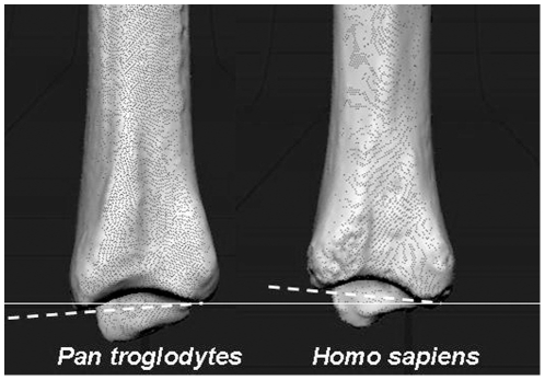
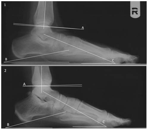
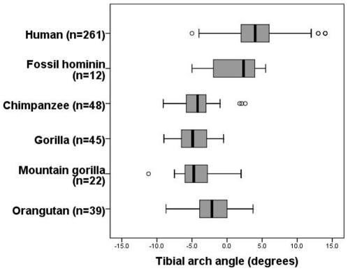
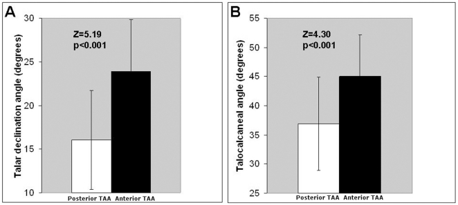
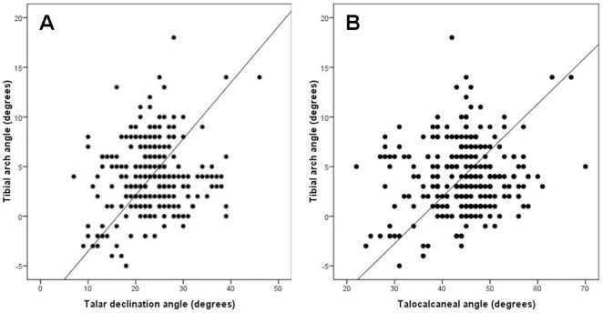
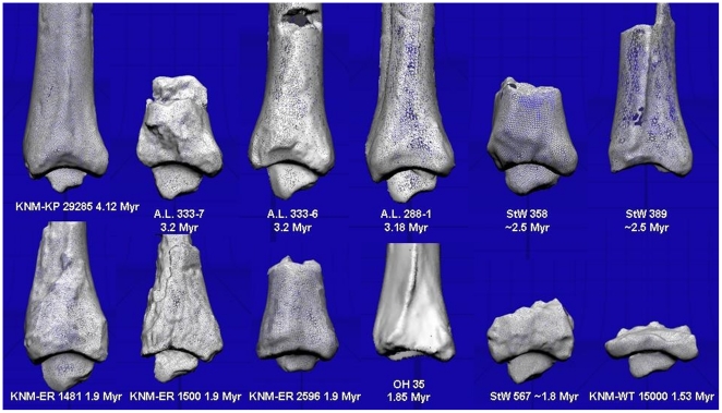
Methodology/principal findings: Here, we present evidence from radiographs of modern humans (n = 261) that the set of the distal tibia in the sagittal plane, henceforth referred to as the tibial arch angle, is related to rearfoot arching. Non-human primates have a posteriorly directed tibial arch angle, while most humans have an anteriorly directed tibial arch angle. Those humans with a posteriorly directed tibial arch angle (8%) have significantly lower talocalcaneal and talar declination angles, both measures of an asymptomatic flatfoot. Application of these results to the hominin fossil record reveals that a well developed rearfoot arch had evolved in Australopithecus afarensis. However, as in humans today, Australopithecus populations exhibited individual variation in foot morphology and arch development, and "Lucy" (A.L. 288-1), a 3.18 Myr-old female Australopithecus, likely possessed asymptomatic flat feet. Additional distal tibiae from the Plio-Pleistocene show variation in tibial arch angles, including two early Homo tibiae that also have slightly posteriorly directed tibial arch angles.

Conclusions/significance: This study finds that the rearfoot arch was present in the genus Australopithecus. However, the female Australopithecus afarensis "Lucy" has an ankle morphology consistent with non-pathological flat-footedness. This study suggests that, as in humans today, there was variation in arch development in Plio-Pleistocene hominins.


Conflict of interest statement

Competing Interests: The authors have declared that no competing interests exist.

Figures

Figure 1
Figure 1. Tibial arch angle in chimpanzee and human.
Humans and non-human primates have distinct tilts to the distal tibia in the sagittal plane. In non-human primates (left, chimpanzee), the anterior rim of the tibia (to the left in the figures) is more inferiorly projecting than the posterior rim, creating a posteriorly directed set to the ankle. In humans (right), the posterior rim is more inferiorly projecting, creating an anteriorly directed set to the ankle. In this image, the thin white line has been drawn through the inferomost projection of the posterior rim of both tibiae and is perpendicular to the long axis of the tibia. The tibial arch angle is formed between this white line and the dotted white line intersecting the anterior rim (negative in chimpanzee; positive in humans).
Figure 2
Figure 2. Measurements taken in this study.
1. A flat foot exhibiting a posteriorly directed tibial set. 2. An arched foot exhibiting an anteriorly directed tibial set. Both 1 and 2: A. Tibial arch angle. B. Calcaneal inclination angle. C. Talar declination angle.
Figure 3
Figure 3. Variation in tibial arch angle in extant apes and fossil hominins.
The tibial arch angle differentiates humans and non-human primates. Mountain gorillas, lowland gorillas, and chimpanzees have statistically indistinguishable tibial arch angles, and orangutans have the least posteriorly directed angle of the great apes. These comparative data do not support the hypothesis that this angle is related to arboreality or hindlimb suspensory abilities. Instead, it is argued in this study that the tibial arch angle is related to rearfoot arching. Humans are quite variable for this measure, and fossil hominins occupy the lower end of the modern human spectrum, though this distribution can be sampled from a modern human population. The median (black bar), interquartile range (box) and overall ranges (whiskers) are illustrated. Outliers defined as 1.5 times the interquartile range are shown as circles.
Figure 4
Figure 4. Relationship between tibial arch angle and rearfoot arching in humans.
Modern humans with a Lucy-like posteriorly directed set to the distal tibia (white bars mean ± sd) have significantly lower talar declination (A) and talocalcaneal angles (B) than modern humans with an anteriorly directed set to the ankle joint (black bars mean ± sd).
Figure 5
Figure 5. Correlation between tibial arch angle and measures of flat foot in humans.
There is a statistically significant positive correlation between the tibial arch angle and two measures of asymptomatic flat-footedness, the talar declination angle (A), and the talocalcaneal angle (B). A regression line generated using reduced major axis regression is drawn in each graph.
Figure 6
Figure 6. Fossil hominin distal tibiae.
Fossil hominin tibiae examined in this study with genus Australopithecus in the top row, and Homo and Paranthropus in the bottom row. All are scans of original fossils with the exception of the three fossils from Hadar, Ethiopia (A.L. fossils), and OH 35. Fossils were 3D laser scanned, scaled to roughly the same size, and presented here to visualize the tibial arch angle. Anterior is to the left, posterior to the right. KNM-KP 29285, A.L. 288-1, and StW 567 have been reversed to reflect the left side. Individual arch angles are presented in Table 2. Notice here the posteriorly directed set to A.L. 288-1, and the slight posteriorly directed set to KNM-KP 29285, StW 567, and KNM-ER 1481. All other fossils show an anteriorly directed set.

Similar articles

Cited by

References

    1. Ker RF, Bennett MB, Bibby SR, Kester RC, Alexander RMcN. The spring in the arch of the human foot. Nature. 1987;325:147–149. - PubMed
    1. Sarrafian SK. Functional characteristics of the foot and plantar aponeurosis under tibiotalar loading. Foot Ankle. 1987;8:4–18. - PubMed
    1. Morton DJ. The Human Foot. New York: Columbia University Press; 1935. 257
    1. Hicks JH. The mechanics of the foot: II. The plantar aponeurosis and the arch. J Anat. 1954;88:25–30. - PMC - PubMed
    1. Harris EJ. The natural history and pathophysiology of flexible flatfoot. Clin Pod Med Surg. 2010;27:1–23. - PubMed