. 2010 Dec 28;5(12):e14421.
doi: 10.1371/journal.pone.0014421.

Unravelling the mystery of stem/progenitor cells in human breast milk

Affiliations

Unravelling the mystery of stem/progenitor cells in human breast milk

Yiping Fan et al. PLoS One. .

Abstract

Background: Mammary stem cells have been extensively studied as a system to delineate the pathogenesis and treatment of breast cancer. However, research on mammary stem cells requires tissue biopsies which limit the quantity of samples available. We have previously identified putative mammary stem cells in human breast milk, and here, we further characterised the cellular component of human breast milk.

Methodology/principal findings: We identified markers associated with haemopoietic, mesenchymal and neuro-epithelial lineages in the cellular component of human breast milk. We found 2.6 ± 0.8% (mean ± SEM) and 0.7 ± 0.2% of the whole cell population (WCP) were found to be CD133+ and CD34+ respectively, 27.8 ± 9.1% of the WCP to be positive for Stro-1 through flow-cytometry. Expressions of neuro-ectodermal stem cell markers such as nestin and cytokeratin 5 were found through reverse-transcription polymerase chain reaction (RT-PCR), and in 4.17 ± 0.2% and 0.9 ± 0.2% of the WCP on flow-cytometry. We also established the presence of a side-population (SP) (1.8 ± 0.4% of WCP) as well as CD133+ cells (1.7 ± 0.5% of the WCP). Characterisation of the sorted SP and non-SP, CD133+ and CD133- cells carried out showed enrichment of CD326 (EPCAM) in the SP cells (50.6 ± 8.6 vs 18.1 ± 6.0, P-value = 0.02). However, culture in a wide range of in vitro conditions revealed the atypical behaviour of stem/progenitor cells in human breast milk; in that if they are present, they do not respond to established culture protocols of stem/progenitor cells.

Conclusions/significance: The identification of primitive cell types within human breast milk may provide a non-invasive source of relevant mammary cells for a wide-range of applications; even the possibility of banking one's own stem cell for every breastfeeding woman.


Conflict of interest statement

Competing Interests: The authors have declared that no competing interests exist.

Figures

Figure 1
Figure 1. Cellular concentration in human breast milk did not vary in relation to the duration of breastfeeding.
Figure 2
Figure 2. RT-PCR on Messenger RNA (mRNA) of milk samples from three individuals.
(a) mRNA of CD133 and CD34 were present in WCP of HBM (Lane 1–3). (b) Osteonectin (ON), alkaline phosphatase (ALP) and osteopontin (OP) (Lane 1–3) as well as (c) musashi-1 (Msi), nestin (NES) and neurofilament-M (NFM) in observed in WCP of HBM (Lane 1–3). (d) Messenger RNA of CK5, 14 and 18 were present in WCP of HBM (Lane 1–3). The negative controls in Lane 4, were MCF-7 for hematopoietic, mesenchymal and neural markers and mononuclear cells in peripheral blood for epithelial cell markers of CK5 and CK14. Positive controls (Lane 5) are cells isolated from umbilical cord blood (a), fetal MSC (b), cells from snap-frozen fetal brain (c) and MCF-7 (d) respectively.
Figure 3
Figure 3. Side-population of HBM.
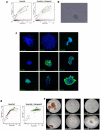
(a) Sorting profile of side-population (SP) and non side-population (NSP) from breast milk. (b) Scatter plots showing no relationship between the percentage of SP and duration of breastfeeding and (c) age of mother. (d) Immunocytochemistry illustrating expression of nestin exclusively only on SP (top left) and expression of CK 18 exclusively on NSP (bottom right).
Figure 4
Figure 4. Culture of SP of positive control.
(a) Flow cytometry profile of MCF-7, a breast cancer cell line from which the side population which was sorted out (b) grew into mammospheres after 10 days with (c) immunocytochemistry of the mammospheres ascertaining their identity. (d) Flow cytometry profile of umbilical cord blood with the side-population sorted out for culture in methylcellulose, (e) confirming the presence of burst forming unit-erythrocyte (BFU-E), colony forming unit-erythrocyte (CFU-E), colony forming unit-macrophage (CFU-M), colony forming unit-granulocyte, erythrocyte, macrophage, megakaryocyte (CFU-GEMM) as well as colony forming unit-granulocyte, macrophage (CFU-GM) in the SP of cord blood.
Figure 5
Figure 5. Mesenchymal Culture of Cells.
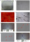
(a, b) Adherent colonies of fetal MSC emerged after low density seeding at 4 cells per cm2. Osteogenic induction of fetal MSC resulted in the deposition of extracellular calcium crystals staining positive with Alizarin red (c) and Von Kossa (d).CFU-F assays of SP and NSP as well as Stro1 positive and negative fractions of HBM did not establish any colonies as shown by the absence of colonies (e, h). WCP (f) and Stro-1 positive cells (g) cultured in D10 remained as non-adherent cells (red arrows) which did not undergo any proliferation in culture.
Figure 6
Figure 6. CD133 Sorting.
(a) Flow cytometry of CD133 staining on cellular component of HBM (i), as compared to the isotype control (ii). (b, c) Three was no relationship between the frequency of CD133 cells and the duration of breastfeeding, nor the age of the mother. (d) CD133+ cells from cord blood formed multi-lineage colonies on CFC assays.

References

    1. Deome KB, Faulkin LJ, Jr, Bern HA, Blair PB. Development of mammary tumors from hyperplastic alveolar nodules transplanted into gland-free mammary fat pads of female C3H mice. Cancer Res. 1959;19:515–520. - PubMed
    1. Gudjonsson T, Villadsen R, Nielsen HL, Ronnov-Jessen L, Bissell MJ, et al. Isolation, immortalization, and characterization of a human breast epithelial cell line with stem cell properties. Genes Dev. 2002;16:693–706. - PMC - PubMed
    1. Dontu G, Abdallah WM, Foley JM, Jackson KW, Clarke MF, et al. In vitro propagation and transcriptional profiling of human mammary stem/progenitor cells. Genes Dev. 2003;17:1253–1270. - PMC - PubMed
    1. Alvi AJ, Clayton H, Joshi C, Enver T, Ashworth A, et al. Functional and molecular characterisation of mammary side population cells. Breast Cancer Res. 2003;5:R1–8. - PMC - PubMed
    1. Shackleton M, Vaillant F, Simpson KJ, Stingl J, Smyth GK, et al. Generation of a functional mammary gland from a single stem cell. Nature. 2006;439:84–88. - PubMed

Publication types