. 2010 Dec 23;5(12):e14426.
doi: 10.1371/journal.pone.0014426.

ATL9, a RING zinc finger protein with E3 ubiquitin ligase activity implicated in chitin- and NADPH oxidase-mediated defense responses

Affiliations

ATL9, a RING zinc finger protein with E3 ubiquitin ligase activity implicated in chitin- and NADPH oxidase-mediated defense responses

Marta Berrocal-Lobo et al. PLoS One. .

Abstract

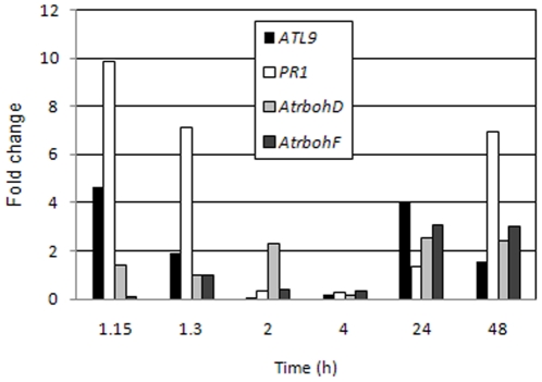
Pathogen associated molecular patterns (PAMPs) are signals detected by plants that activate basal defenses. One of these PAMPs is chitin, a carbohydrate present in the cell walls of fungi and in insect exoskeletons. Previous work has shown that chitin treatment of Arabidopsis thaliana induced defense-related genes in the absence of a pathogen and that the response was independent of the salicylic acid (SA), jasmonic acid (JA) and ethylene (ET) signaling pathways. One of these genes is ATL9 ( = ATL2G), which encodes a RING zinc-finger like protein. In the current work we demonstrate that ATL9 has E3 ubiquitin ligase activity and is localized to the endoplasmic reticulum. The expression pattern of ATL9 is positively correlated with basal defense responses against Golovinomyces cichoracearum, a biotrophic fungal pathogen. The basal levels of expression and the induction of ATL9 by chitin, in wild type plants, depends on the activity of NADPH oxidases suggesting that chitin-mediated defense response is NADPH oxidase dependent. Although ATL9 expression is not induced by treatment with known defense hormones (SA, JA or ET), full expression in response to chitin is compromised slightly in mutants where ET- or SA-dependent signaling is suppressed. Microarray analysis of the atl9 mutant revealed candidate genes that appear to act downstream of ATL9 in chitin-mediated defenses. These results hint at the complexity of chitin-mediated signaling and the potential interplay between elicitor-mediated signaling, signaling via known defense pathways and the oxidative burst.


Conflict of interest statement

Competing Interests: The authors have declared that no competing interests exist.

Figures

Figure 1
Figure 1. ATL9 structure and sequence alignment between ATL family members.
A) Schematic diagram of ATL9 protein structure. The protein has an N terminal region (cytosolic), containing a signal peptide (gray); two transmembrane domains (Tm1 and Tm2, in black); and a C3HC3 RING Zinc-finger domain (dark gray), a PEST sequence and a coiled coil domain that extends into the ER lumen (white). B) Alignment of ATL9 RING Zinc-finger domain amino acid sequence with the other ATL family members implicated in defense responses in plants. Asterisks indicate conserved cysteines; conserved amino acids are indicated in black, residues conserved in more than an 87.5% are highlighted in gray. The consensus sequence for this group of RING zinc-fingers is: C-X2-CL-X-E-X7-R-X2-P-X-C-X-H-X-FH-X2-C-X-D-X-W-X6-CP-X-C, where X is any amino acid. Induction of the corresponding genes is indicated as: C (chitin), F (flagellin), P (pathogens), Av (Avr9), BTH (benzothaidiazole), ACC (1-aminocyclopropane-1-carboxylic acid), CWP (cell wall protein fraction elicitor), (+) indicates mutants in these genes have been tested for altered pathogen response.
Figure 2
Figure 2. Influence of SA-, JA- and ET-mediated pathways on ATL9 expression measured by qRT-PCR.
A) Induction of ATL9 30 minutes after chitin treatment in Col-0 wild-type plants and in the ein2-5, jar1 and sid2-1 mutants. B) Induction of ATL9 (black), PR1 (light gray) and PDF1.2 (dark gray) in Col-0 plants after treatment with chitin (CSC), 50 µM ACC, 5 µM JA or 0.5 mM SA. Data represent the ratio between mock and treated plants of three independent biological samples. The inset shows relative expression of ATL9 transcript compared to the actin control (RE/Actin).
Figure 3
Figure 3. Susceptibility of atl9 mutants to Golovinomycetes cichoracearum and hydrogen peroxide production.
A) Quantification of Golovinomyces cichoracearum growth on Col-0 plants, three different atl9 T-DNA insertional mutants (atl9-1-3), 35S:ATL9, Kas-1, AtrbohD, AtrbohF, AtrbohD/F and the SA-compromised mutant sid2-1. The number of conidiophores per colony (c/c) were counted 6 dpi. Inoculations were performed at a low density (5–10 conidia/mm2; n = 36 plants). Data values represent one of at least three independent experiments with similar results. B) Production of hydrogen peroxide on plants infected with G. cichoracearum. Cleared leaves of infected plants were stained with trypan blue which stains the fungal hyphae and conidiophores on the leaf surface. Secondary staining with DAB was performed to indicate areas of hydrogen peroxide production (indicated by peroxidase activity). a, Kas-1, b, Col-0, c, atl9-1, d, AtrbohD, f, AtrbohD/F, g, sid2-1. Arrows indicate fungal penetration points in epidermal cells with detected hydrogen peroxide production. The images were taken at a final magnification of 40x. Bars.- 5 µm. C) Quantification of hydrogen peroxide production in mutants. The intensity of DAB staining was quantified at each inoculated plant and compared to the corresponding uninoculated control to obtain a relative DAB signal. The quantification was performed with the aid of the program Image J.
Figure 4
Figure 4. The basal expression of ATL9 and its induction by chitin depends on NADPH oxidases.
Plants were treated with chitin for 30 minutes and harvested for analysis. Expression levels of ATL9 were measured by qRT-PCR. The inset shows the relative expression of ATL9 transcript compared to actin (RE/Actin) in control plants. Data represent the ratio of expression between mock and treated plants of three independent biological samples.
Figure 5
Figure 5. Relative expression levels of selected genes at various time points after inoculation with Golovinomyces cichoracearum.
Col-0 plants were inoculated with G. cichoracearum at high concentration. qRT-PCR data represent the ratio between mock and treated plants of three independent biological samples.
Figure 6
Figure 6. ATL9 is capable of mediating protein ubiquitination in a E2-dependent manner.
The complete in vitro ubiquitination assay (lane 1) contained recombinant yeast E1 enzyme, recombinant 6x histidine-tagged Arabidopsis E2 enzyme UBC8, GST-tagged ATL9 and ubiquitin. Omission of AtUBC8 (lane 2), GST-ATL9 (lane 3) or ubiquitin (lane 4) from the assay resulted in a loss of protein polyubiquitination as indicated by a lack of the ubiquitinated proteins compared to the complete assay (lane 1). Ubiquitinated proteins were visualized by western blot analysis using anti-ubiquitin antibodies. * Indicate non-specific cross-reactive proteins. ♦ Represent AtUBC8-Ub(n). Molecular weight markers (kDa) are shown to the left of the blot.
Figure 7
Figure 7. The ATL9 protein localizes to the ER.
A–C) Confocal images of leaf epidermal cells in transgenic Arabidopsis plants expressing ATL9p:ATL9:GFP showing protein localization to the endoplasmic reticulum. B) Confocal image of a leaf trichome showing ATL9p:ATL9:GFP localizing to the ER. D–F) Co-localization of the 35S:ATL9:GFP fusion and ER-rk marker. Onion epidermal cells were co-bombarded with 35S:ATL9:GFP and ER-rk and visualized using fluorescence microscopy. D) 35S:ATL9:GFP. E) ER-rk (mCherry). F) GFP/mCherry overlay. Bars: 3 µm (A,B), 1 µm (C), 10 µm (D-F).
Figure 8
Figure 8. Microarray analysis of atl9 mutant after chitin treatment.
A) Hierarchical cluster of ratio values (relative to the water control treatment) of 16,530 genes analyzed in Col-0 wild-type and atl9 plants treated with crab shell chitin (CSC) for 30 minutes. Each gene is represented by a single row and each column represents an individual treatment. Red represents up-regulated genes; blue, down-regulated genes; and yellow, genes with no change in expression. Groups of genes expressed differentially are delineated with rectangles. Three genes (black arrows) were changing due to interactions between treatment and genotype and one gene (red arrow) was genotype interaction specific. B) Venn diagram representation of results from hierarchical clustering. A total of 4,375 genes were differentially expressed between genotypes and treatments. The statistic used for clustering was two-way ANOVA. Genotype and treatment groups were analyzed using a p-value of 0.5 with p-value >0.5 =  not significant; p-value <0.5 =  significant.
Figure 9
Figure 9. Proposed role of ATL9 in chitin-NADPH mediated innate immunity.
1. Recognition of chitin released from fungal cell wall by LysM-RLK1 receptor. 2. Increase of intra-cellular calcium leading to the activation of the NADPH oxidases AtrbohD and F at the membrane initiating the production of ROS and activation of a MAPK cascade(s). 3. Induction of ATL9 and insertion into the ER membrane. We propose that ATL9 is involved tagging an inhibitor of plant defense for degradation by the proteosome 4. Defense response to fungal pathogen is activated after inhibitor degradation. 5. The ATL9 protein is rapidly degraded in the cell by either the proteosome pathway or a calcium-mediated pathway (similar to the mammalian protein calpain).

References

    1. Osbourn AE. Preformed antimicrobial compounds and plant defense against fungal attack. Plant Cell. 1996;8:1821–1831. - PMC - PubMed
    1. Flor HH. Current status of the gene-for-gene concept. Annu Rev Phytopathol. 1971;9:275–296.
    1. Gomez-Gomez L. Plant perception systems for pathogen recognition and defence. Molecular Immunology. 2004;41:1055–1062. - PubMed
    1. Libault M, Wan JR, Czechowski T, Udvardi M, Stacey G. Identification of 118 Arabidopsis transcription factor and 30 ubiquitin-ligase genes responding to chitin, a plant-defense elicitor. Molecular Plant-Microbe Interactions. 2007;20:900–911. - PubMed
    1. Navarro L, Zipfel C, Rowland O, Keller I, Robatzek S, et al. The transcriptional innate immune response to flg22. interplay and overlap with Avr gene-dependent defense responses and bacterial pathogenesis. Plant Physiology. 2004;135:1113–1128. - PMC - PubMed

Publication types

MeSH terms

LinkOut - more resources