. 2010 Dec 23;5(12):e15330.
doi: 10.1371/journal.pone.0015330.

Circadian disruption accelerates tumor growth and angio/stromagenesis through a Wnt signaling pathway

Affiliations

Circadian disruption accelerates tumor growth and angio/stromagenesis through a Wnt signaling pathway

Yoshihiro Yasuniwa et al. PLoS One. .

Abstract

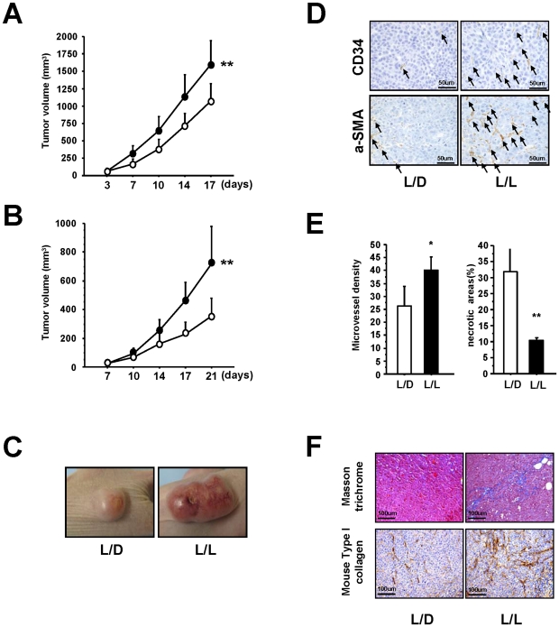
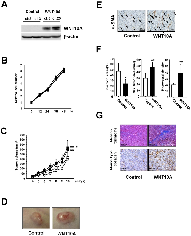
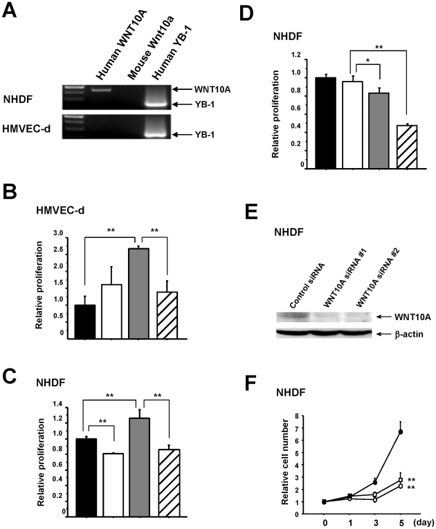
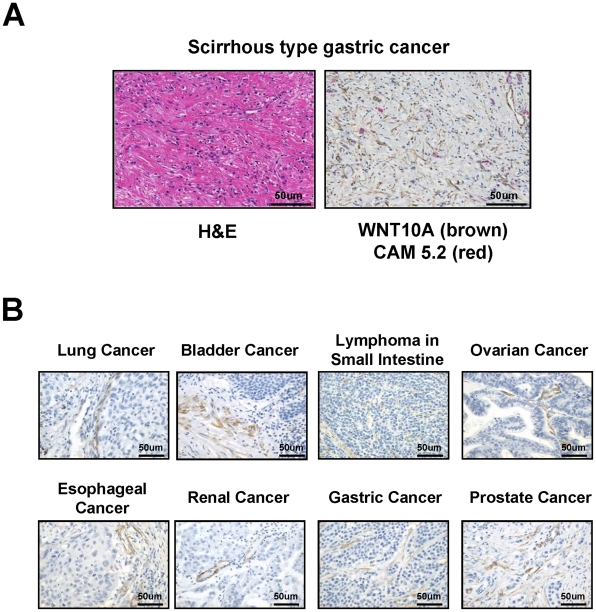
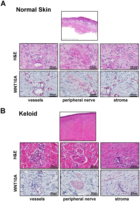
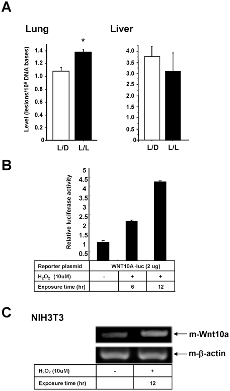
Epidemiologic studies show a high incidence of cancer in shift workers, suggesting a possible relationship between circadian rhythms and tumorigenesis. However, the precise molecular mechanism played by circadian rhythms in tumor progression is not known. To identify the possible mechanisms underlying tumor progression related to circadian rhythms, we set up nude mouse xenograft models. HeLa cells were injected in nude mice and nude mice were moved to two different cases, one case is exposed to a 24-hour light cycle (L/L), the other is a more "normal" 12-hour light/dark cycle (L/D). We found a significant increase in tumor volume in the L/L group compared with the L/D group. In addition, tumor microvessels and stroma were strongly increased in L/L mice. Although there was a hypervascularization in L/L tumors, there was no associated increase in the production of vascular endothelial cell growth factor (VEGF). DNA microarray analysis showed enhanced expression of WNT10A, and our subsequent study revealed that WNT10A stimulates the growth of both microvascular endothelial cells and fibroblasts in tumors from light-stressed mice, along with marked increases in angio/stromagenesis. Only the tumor stroma stained positive for WNT10A and WNT10A is also highly expressed in keloid dermal fibroblasts but not in normal dermal fibroblasts indicated that WNT10A may be a novel angio/stromagenic growth factor. These findings suggest that circadian disruption induces the progression of malignant tumors via a Wnt signaling pathway.


Conflict of interest statement

Competing Interests: The authors have declared that no competing interests exist.

Figures

Figure 1
Figure 1. Effect of photoperiod manipulation on the growth of human HeLa cell or PC3 cell tumors.
(A) Hela cell tumors. Volume of the subcutaneous xenografts in nude mice housed in either L/L (closed circle; n = 16) or L/D (open circle; n = 16) conditions. After 17 days of growth, L/L tumors were significantly larger than L/D tumors (F from repeated measure ANOVA = 12.276, **P<0.01). (B) PC3 cell tumors. Volume of the subcutaneous xenografts in nude mice housed in either L/L (n = 8) or L/D (n = 8) conditions. A significant reduction in volume is seen in the L/D tumors compared with the L/L tumors (F from repeated measure ANOVA  = 18.360, **P<0.01). (C) Representative photograph of L/L and L/D tumors showing the obvious difference in size. (D) Immunohistochemical analysis of CD34 positive (CD34+) cells and aSMA positive (aSMA+) cells in the L/L and L/D tumors. Increased numbers of CD34+ and aSMA+ (black arrows) are clearly visible in the L/L tumors. (E) The number of microvessels was quantified using the number of CD34+ cells. An increase in microvessel density (vessels in 10 viewing fields; n = 3 per group, *P<0.05), is accompanied by a decrease in necrosis (n = 3 per group **P<0.01). (F) Representative photographs showing masson trichrome staining of the expanded extracellular matrix and immunohistochemical analysis of mouse Type I collagen in the L/D and L/L tumors.
Figure 2
Figure 2. WNT10A was upregulated in L/L tumors.
(A) RT-PCR of the relevant gene transcripts was carried out based on the results of the DNA microarray analysis. Human WNT10A (h-WNT10), mouse Wnt10a (m-Wnt10a) and ANGPL4 were upregulated in L/L mice tumors. RB1 was downregulated. There was no difference in the expression levels of VEGF-A, VEGF-B and YB-1 between L/L and L/D tumors. Human β-actin (h-β-actin) and mouse β-actin (m-β-actin) were used as internal control. The cycle number is 30 for all semi-quantitative RT-PCR except h-WNT10A. Nested PCR technique to investigate the expression of human WNT10A in tumors. The cycle number of 1st PCR is 30 and that of 2nd nested PCR is 35. (B) Immunohistochemical analysis of WNT10A in L/L and L/D mice tumors. The arrows indicate tumor blood vessels.
Figure 3
Figure 3. WNT10A functions as an angio/stromagenetic growth factor in vivo xenograft models.
(A) Establishment of a stable WNT10A-overexpressing cell line. (B), (C) The growth rate of these stable cell lines (B) in vitro and (C) in vivo. Two control cell lines (cl:2; open circle, cl:3; open square) and two stable WNT10A-overexpressing cell lines (cl:6; closed circle, cl:25, closed square) were used. **P<0.01 compared with the control cl:2 group and #P<0.05 compared with the control cl:3 group using Scheffe's test. n = 8 per groups. (D) Representative photograph WNT10A-overexpressing tumors in nude mice illustrating their hypervascular nature. (E) Immunostaining of tumors with an anti-aSMA antibody. Increased numbers of aSMA+ cells (black arrows) are clearly visible in WNT10A-overexpressing tumors. (F) Reduced areas of tissue necrosis in WNT10A-overexpressing tumors are accompanied by increased tumor size and increased microvessel density (n = 4 or 6 per group, *P<0.05 and **P<0.01). Microvessel density was quantified using the number of aSMA+ cells (G) Masson trichrome staining showing expansion of the extracellular matrix and immunohistchemical analysis of mouse Type I collagen in the control and WNT10A-overexpressing tumors.
Figure 4
Figure 4. WNT10A is expressed in fibroblast cells and functions as an angio/stromagenesis growth factor in vitro.
(A) RT-PCR for human WNT10A mRNA in NHDF and HMVEC-d cells. Human YB-1 was used as a positive control and mouse Wnt10a was used as a negative control. (B), (C) WNT10A-dependent growth of HMVEC-d and NHDF cells. For the proliferation assays using BrdU incorporation, HMVEC-d (NHDF) cells were cultured in conditioned medium (CM) with or without WNT10A antibody for 24 hours (black bar; Control-CM, white bar; Control-CM + WNT10A antibody 5 µg/ml, gray bar; WNT10A-CM, slash bar; WNT10A-CM + WNT10A antibody 5 µg/ml). **P<0.01. n = 3 per groups. (D) WNT10A-dependent autocrine growth of NHDF cells. NHDF cells were cultured with the recommended medium (RM) with or without WNT10A antibody (black bar; RM, white bar; RM + IgG 5 µg/ml, gray bar; RM + WNT10A antibody 2 µg/ml, slash bar; RM + WNT10A antibody 5 µg/ml). *P<0.05 and **P<0.01. n = 3 per groups. (E) Complete knockdown of WNT10A expression in NHDF cells is achieved using the two siRNAs against WNT10A. Whole-cell extracts (100 µg) were subjected to SDS-PAGE, and Western blotting analysis was performed using the indicated antibodies. (F) Knockdown of WNT10A suppresses the growth of NHDF cells (Control siRNA; closed circle, WNT10A siRNA #1; open square, WNT10A siRNA #2; open circle). **P<0.01 compared with the control siRNA group.
Figure 5
Figure 5. Immunohistochemical analysis of WNT10A expression in various human cancer specimens.
(A) H&E and double immunohistochemical staining of scirrhous-type signet-ring carcinoma cells. 3,3′-Diaminobenzidine (DAB) was used as chromogen for WNT10A staining (brown color) and Vulcan fast red was used for cytokeratin CAM 5.2 staining (red color). Anti-cytokeratin CAM 5.2 was used for staining of signet-ring cell carcinoma cells. (B) WNT10A expresses cancer stroma cells in various human cancer specimens. 3,3′-Diaminobenzidine (DAB) was used as chromogen for WNT10A staining (brown color).
Figure 6
Figure 6. WNT10A is expressed in stromal cells of keloid tissue.
H&E and anti-WNT10A antibody staining in (A) normal skin and (B) keloid tissue. WNT10A was expressed around the vessels and in the peripheral nerve cells of normal skin, but not in stromal cells. In contrast, WNT10A was expressed around vessels, in peripheral nerve cells and strongly in stromal cells of keloid tissue. Vulcan fast red was used for WNT10A staining (red color).
Figure 7
Figure 7. WNT10A is induced by oxidative stress.
(A) 8-hydroxydeoxyguanosine levels are significantly increased in the lung tissues of L/L compared with L/D mice (*P<0.05). (B) Reporter assays. The promoter activity of the WNT10A gene was measured using a luciferase system after the addition of hydrogen peroxide. 42 hours after transfection (exposure time 6 hr) or 36 hours after transfection (exposure time 12 hr) of the reporter plasmid into PC3 cells, cells were treated with 10 µM of hydrogen peroxide. Luciferase activities were assayed after 48 hours of transfection. The results shown are normalized against protein concentrations measured using the Bradford method and are representative of at least three independent experiments. (C) Induction of mouse Wnt10a transcripts by oxidative stress. NIH3T3 cells were treated with or without H2O2 (10 µM) for 12 hours. Total RNAs were assayed by semi- quantitative RT-PCR. Mouse β-actin was used for internal control.

Similar articles

Cited by

References

    1. Rajaratnam SM, Arendt J. Health in a 24-h society. Lancet. 2006;358:999–1005. - PubMed
    1. Navara KJ, Nelson RJ. The dark side of light at night: physiological, epidemiological, and ecological consequences. J Pineal Res. 2007;43:215–24. - PubMed
    1. Fu L, Lee CC. The circadian clock: pacemaker and tumour suppressor. Nat Rev Cancer. 2003;3:350–61. - PubMed
    1. Antoni MH, Lutgendorf SK, Cole SW, Dhabhar FS, Sephton SE, et al. The influence of bio-behavioural factors on tumour biology: pathways and mechanisms. Nat Rev Cancer. 2006;6:240–8. - PMC - PubMed
    1. Hansen J. Risk of breast cancer after night- and shift work: current evidence and ongoing studies in Denmark. Cancer Causes Control. 2006;17:531–7. - PubMed

Publication types