. 2010 Dec 22;5(12):e15773.
doi: 10.1371/journal.pone.0015773.

Detection and functional characterization of a 215 amino acid N-terminal extension in the Xanthomonas type III effector XopD

Affiliations

Detection and functional characterization of a 215 amino acid N-terminal extension in the Xanthomonas type III effector XopD

Joanne Canonne et al. PLoS One. .

Retraction in

Abstract

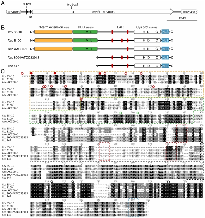
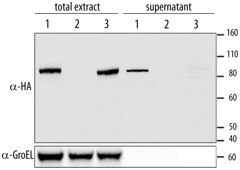
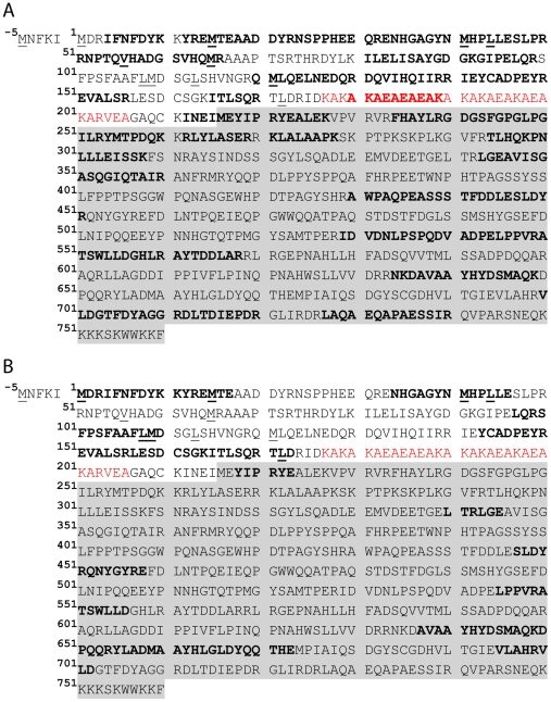
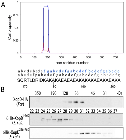
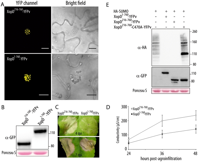
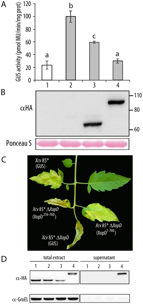
During evolution, pathogens have developed a variety of strategies to suppress plant-triggered immunity and promote successful infection. In Gram-negative phytopathogenic bacteria, the so-called type III protein secretion system works as a molecular syringe to inject type III effectors (T3Es) into plant cells. The XopD T3E from the strain 85-10 of Xanthomonas campestris pathovar vesicatoria (Xcv) delays the onset of symptom development and alters basal defence responses to promote pathogen growth in infected tomato leaves. XopD was previously described as a modular protein that contains (i) an N-terminal DNA-binding domain (DBD), (ii) two tandemly repeated EAR (ERF-associated amphiphillic repression) motifs involved in transcriptional repression, and (iii) a C-terminal cysteine protease domain, involved in release of SUMO (small ubiquitin-like modifier) from SUMO-modified proteins. Here, we show that the XopD protein that is produced and secreted by Xcv presents an additional N-terminal extension of 215 amino acids. Closer analysis of this newly identified N-terminal domain shows a low complexity region rich in lysine, alanine and glutamic acid residues (KAE-rich) with high propensity to form coiled-coil structures that confers to XopD the ability to form dimers when expressed in E. coli. The full length XopD protein identified in this study (XopD(1-760)) displays stronger repression of the XopD plant target promoter PR1, as compared to the XopD version annotated in the public databases (XopD(216-760)). Furthermore, the N-terminal extension of XopD, which is absent in XopD(216-760), is essential for XopD type III-dependent secretion and, therefore, for complementation of an Xcv mutant strain deleted from XopD in its ability to delay symptom development in tomato susceptible cultivars. The identification of the complete sequence of XopD opens new perspectives for future studies on the XopD protein and its virulence-associated functions in planta.


Conflict of interest statement

Competing Interests: The authors have declared that no competing interests exist.

Figures

Figure 1
Figure 1. In silico analysis of the xopD locus.
(A) The xopD locus in Xcv 85-10 is shown (the genome interval for the Xcv 85-10 shown sequence and the xopD gene are respectively: 486209–489200 and 486544–488826). Open arrows indicate ORFs and filled arrows show promoter elements, such as the PIP box and the −10 sequence. The position of XopD translation start annotated in the public databases is indicated by an asterisk. (B) Schematic representation of XopD functional domains in Xcv 85-10, in comparison to its known protein homologues. The N-terminal extension, essential V and L residues in the DBD, tandemly repeated EAR motifs, conserved catalytic residues in the cysteine protease domain, and NLS motif are shown. (C) Sequence alignment of XopD from Xcv 85-10, hypothetical protein from Xcc B100 (YP_001902662), virulence protein from Xcc 8004 (AAY48282) or Xcc ATCC 33913 (AAM42168), peptidase C48 SUMO from Acidovorax avenae (EFA39722) and XopD from Xcc 147. The figure shows the longest ORF possible for all proteins. For XopD from Xcv, the first shown M residue corresponds to a UUG codon, not conserved among Xcc B100 and A. avenae coding sequences. The M residue previously annotated as the starting amino acid of the XopD protein is indicated by a red arrowhead. Putative translation starts situated in frame and upstream the previously annotated starting M are also indicated as follows: translation starts conserved among Xcv 85-10, Xcc B100 and Aea are indicated with a full red dot, otherwise, they appear indicated by an empty red dot. Yellow box: putative N-terminal extension; green box: DNA-binding domain (V and L residues in the helix-loop-helix domain essential for maximal DNA-binding are indicated by an empty green dot); red boxes: tandemly-repeated EAR motifs [L/FDNLL/F(X)P] ; black box: SUMO protease domain (H, D and C catalytic core residues are indicated by a black empty dot); blue box: NLS. Gray and black highlighting of amino acids indicates, respectively, 70–80% and 90–100% of similarity.
Figure 2
Figure 2. Expression analysis of XopD.
Strains 85* (XopD-HA) (1), 85* (2) and 85* ΔhrcV (XopD-HA) (3) were incubated in MOKA rich medium (total extract, left) or secretion medium (supernatant, right). Total protein extracts (10-fold concentrated) and TCA-precipitated filtered supernatants (200-fold concentrated) were analyzed by immunoblotting using anti-HA antibodies (upper panel) to detect the presence of XopD, or anti-GroEL antibodies (lower panel) to show that bacterial lysis had not occurred.
Figure 3
Figure 3. Analysis of the XopD protein sequence by mass spectrometry.
XopD216-760 protein sequence is shadowed. All possible translation starts situated in frame and upstream the annotated M216 are underlined. Peptides identified by Nano LC/ESI MS/MS analysis following trypsin (A) or V8 protease (B) digestion of the purified XopD protein are shown in bold. The 30 amino acid low complexity KAE-rich region is indicated in red.
Figure 4
Figure 4. XopD1-760, but not XopD216-760, is able to dimerize.
(A) Multicoil (http://groups.csail.mit.edu/cb/multicoil/cgi-bin/multicoil.cgi) prediction on XopD1-760 sequence. XopD amino acid sequence (amino acids 1 to 760) was analyzed for its propensity to form coiled-coil structures. A region comprising amino acid residues 168–202 showed high probability to form such coiled-coil structures. Furthermore, this coiled-coil region is likely to be involved in dimer formation (blue line), rather than trimer (red line) or other oligomeric structures. The (abcdefg) positions of the hepta-repeat in the coiled-coil region are indicated above the amino acid sequence. Above it, the relative positions of the hepta-repeat in the opposite chain within the putative dimer are indicated in blue. (B) Gel filtration analysis of HA-tagged XopD expressed in Xcv, and XopD1-760 and XopD216-760 expressed in E. coli as 6xHis-tagged fusions. Protein extracts were subjected to gel filtration chromatography on a Superdex S-200 column. 0.4 ml fractions were collected and aliquots were analyzed by Western blot. Fraction numbers of the elution profile re indicated by the numbers between the gels. The molecular mass estimated for each fraction (in kDa) is given at the top.
Figure 5
Figure 5. Characterization of XopD1-760 expression and function in N. benthamiana.
(A) Confocal images of epidermal cells of N. benthamiana leaves expressing YFPv-tagged XopD216-760 and XopD1-760 36 hours after agroinfiltration. Bright field images are shown on the right. Bars  = 15 µm. (B) Western blot analysis using an anti-GFP antibody shows expression of YFPv-tagged XopD216-760 and XopD1-760 constructs, 36 hours after agroinfiltration. Ponceau S staining of the membrane illustrates equal loading. (C) Cell death development 4 (upper panel) and 7 (lower panel) days after agroinfiltration of YFPv-tagged XopD216-760 (left) and XopD1-760 (right) constructs. (D) Cell death was quantified by measuring electrolyte leakage in N. benthamiana leaves expressing YFPv-tagged XopD216-760 (open circles) and XopD1-760 (filled circles) at the indicated time points after agroinfiltration. Mean and SEM values are calculated from 3 independent experiments (8 replicates/experiment). The statistical significance in mean conductivity values obtained with leaves expressing XopD216-760 or XopD1-760 was assessed by using a Student's t test (P value <10−5). (E) SUMO-conjugates detected by Western blot analysis using an anti-HA antibody 36 hours after agroinfiltration of the indicated constructs. Expression of XopD proteins was revealed using an anti-GFP antibody. Ponceau S staining of the membrane illustrates equal loading.
Figure 6
Figure 6. In planta analysis of XopD1-760-mediated virulence functions.
(A) Transactivation of the PR1 promoter after SA treatment in transient assays in N. benthamiana. Leaves were inoculated with A. tumefaciens carrying a 35S:PR1p-GUS fusion either alone (lanes 1, 2) or together with HA-tagged XopD216-760 (lane 3) or XopD1-760 (lane 4). 18 hours after agroinfiltration, leaves were mock-treated (white bar) or treated with 2 mM SA (grey bars). Fluorimetric GUS assays in leaf discs were performed 12 hours later. Mean values and SEM values were calculated from the results of four independent experiments, with four replicates per experiment. Statistical differences according to a Student's t test P value <0.05 are indicated by letters. MU, methylumbelliferone. (B) Western blot analysis showing expression of HA-tagged XopD216-760 and XopD1-760. Ponceau S staining illustrates equal loading. (C) Susceptible Pearson tomato leaves were inoculated with Xcv 85* or Xcv 85* ΔxopD, expressing an HA-tagged GUS control, Xcv 85* ΔxopD expressing HA-tagged XopD216-760 or Xcv 85* ΔxopD expressing HA-tagged XopD1-760. Inoculation was performed with bacterial suspensions of 1×10⁵ cfu/ml. Representative symptoms observed 10 dpi are shown. Similar phenotypes were observed in four independent experiments. (D) Strains Xcv 85* expressing a GUS control (1) and 85* ΔxopD expressing either a GUS control (2), XopD216-760 (3) or XopD1-760 (4) were incubated in MOKA rich medium (total extract, left) or secretion medium (supernatant, right). Total protein extracts (10-fold concentrated) and TCA-precipitated filtered supernatants concentrated (200-fold concentrated) were analysed by immunoblotting using anti-HA antibodies (upper panel) to detect the presence of GUS, XopD216-760 and XopD1-760, or anti-GroEL antibodies (lower panel) to show that bacterial lysis had not occurred.

References

    1. Zipfel C. Pattern-recognition receptors in plant innate immunity. Curr Opin Immunol. 2008;20:10–16. - PubMed
    1. Jones JD, Dangl JL. The plant immune system. Nature. 2006;444:323–329. - PubMed
    1. Loake G, Grant M. Salicylic acid in plant defence: the players and protagonists. Curr Opin Plant Biol. 2007;10:466–472. - PubMed
    1. Tao Y, Xie Z, Chen W, Glazebrook J, Chang HS, et al. Quantitative Nature of Arabidopsis Responses during Compatible and Incompatible Interactions with the Bacterial Pathogen Pseudomonas syringae. Plant Cell. 2003;15:317–330. - PMC - PubMed
    1. Torres MA, Dangl JL, Jones JD. Arabidopsis gp91phox homologues AtrbohD and AtrbohF are required for accumulation of reactive oxygen intermediates in the plant defense response. Proceedings of The National Academy of Sciences of USA. 2002;99:517–522. - PMC - PubMed

Publication types