. 2010 Dec 23;6(12):e1001234.
doi: 10.1371/journal.ppat.1001234.

Role for sumoylation in systemic inflammation and immune homeostasis in Drosophila larvae

Affiliations

Role for sumoylation in systemic inflammation and immune homeostasis in Drosophila larvae

Indira Paddibhatla et al. PLoS Pathog. .

Abstract

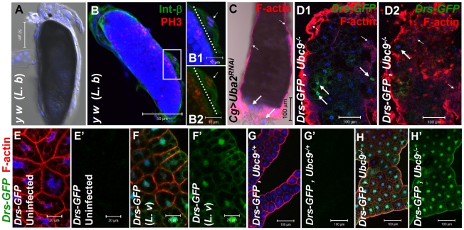
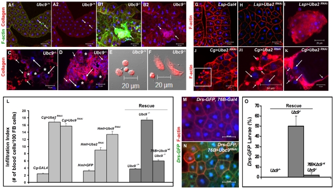
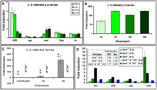
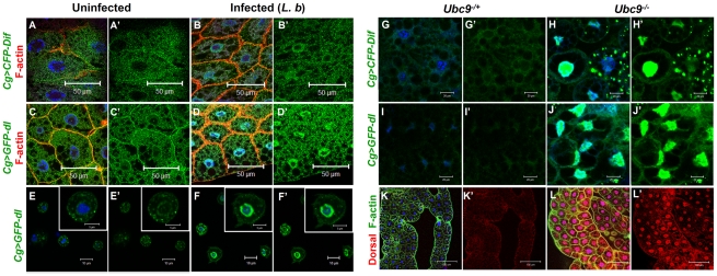
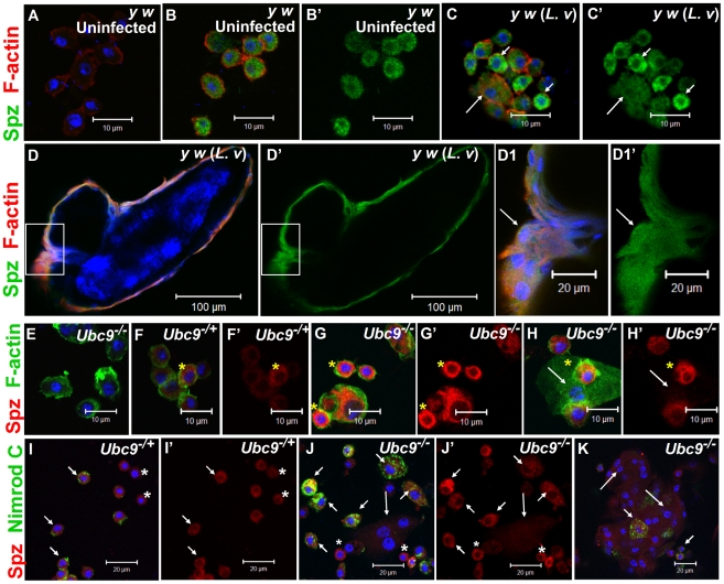
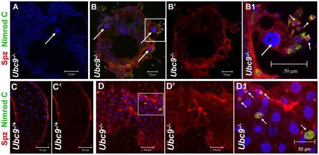
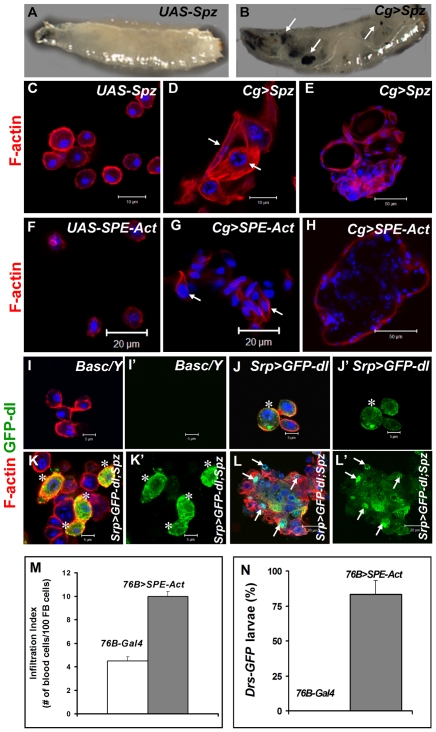
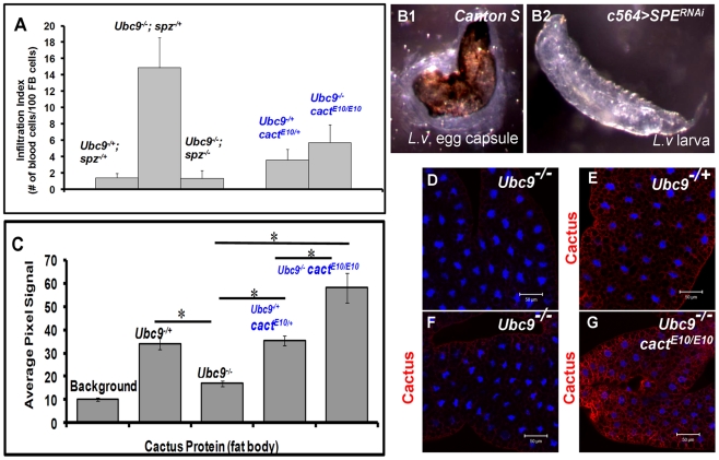
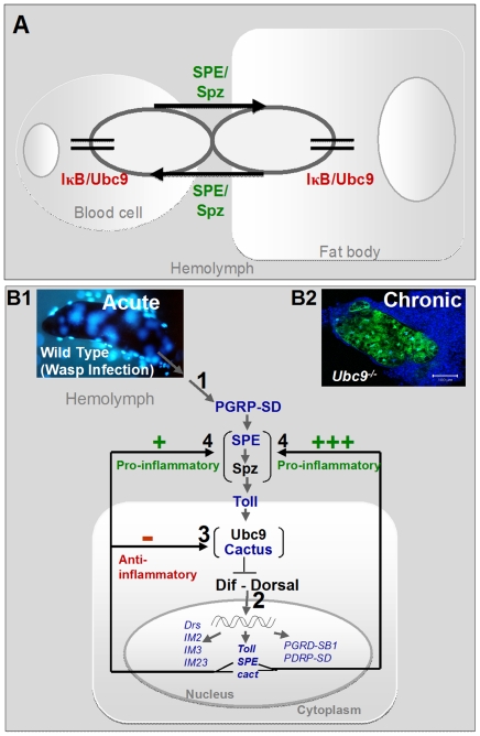
To counter systemic risk of infection by parasitic wasps, Drosophila larvae activate humoral immunity in the fat body and mount a robust cellular response resulting in encapsulation of the wasp egg. Innate immune reactions are tightly regulated and are resolved within hours. To understand the mechanisms underlying activation and resolution of the egg encapsulation response and examine if failure of the latter develops into systemic inflammatory disease, we correlated parasitic wasp-induced changes in the Drosophila larva with systemic chronic conditions in sumoylation-deficient mutants. We have previously reported that loss of either Cactus, the Drosophila (IκB) protein or Ubc9, the SUMO-conjugating enzyme, leads to constitutive activation of the humoral and cellular pathways, hematopoietic overproliferation and tumorogenesis. Here we report that parasite infection simultaneously activates NF-κB-dependent transcription of Spätzle processing enzyme (SPE) and cactus. Endogenous Spätzle protein (the Toll ligand) is expressed in immune cells and excessive SPE or Spätzle is pro-inflammatory. Consistent with this function, loss of Spz suppresses Ubc9⁻ defects. In contrast to the pro-inflammatory roles of SPE and Spätzle, Cactus and Ubc9 exert an anti-inflammatory effect. We show that Ubc9 maintains steady state levels of Cactus protein. In a series of immuno-genetic experiments, we demonstrate the existence of a robust bidirectional interaction between blood cells and the fat body and propose that wasp infection activates Toll signaling in both compartments via extracellular activation of Spätzle. Within each organ, the IκB/Ubc9-dependent inhibitory feedback resolves immune signaling and restores homeostasis. The loss of this feedback leads to chronic inflammation. Our studies not only provide an integrated framework for understanding the molecular basis of the evolutionary arms race between insect hosts and their parasites, but also offer insights into developing novel strategies for medical and agricultural pest control.


Conflict of interest statement

The authors have declared that no competing interests exist.

Figures

Figure 1
Figure 1. Inflammatory responses in the fly larvae.
(A) Melanized wasp (L. boulardi) egg encapsulated by blood cells. (B) Integrin β PS (green) is expressed in most blood cells of the capsule. Some blood cells are mitotically active, where the phospho-histone H3 (PH 3, red) signal overlaps with DNA (B1, B2). Inset shows higher magnification of wasp embryo to which integrin β PS-positive (blood cells, [52]) and some PH 3-positive blood cells adhere (short arrows in B1–B2). PH 3-positive cells are also detected within the developing wasp embryo. Broken line shows the wasp embryo/host capsule interface (B1, B2). (C) Melanized microtumor showing fat body encapsulated by blood cells in Cg>Uba2RNAi larva. Microtumors are structures larger than aggregates of more than 50 blood cells, but smaller than 1 mm3 in volume. Long arrows point to fat body and short arrows to blood cells adhering to the fat body. Blood cells are diploid and significantly smaller than the larger, endopolyploid fat body cells. (D1, D2) Confocal sections of a microtumor (D1, tumor interior; D2 tumor periphery) showing blood cells (short arrows) infiltrating fat body cells. Some fat body cells show constitutive Drs-GFP expression (long arrows). (E–H) Drs-GFP expression in fat body before (E, E'), or after (F, F') wasp (L. victoriae) infection. (G–H) Drs-GFP expression in heterozygous (G, G'), and Ubc9 (H, H') fat body. Panels E', F', G' and H' show Drs-GFP transgene expression alone. (A–H) Cells counterstained for DNA with Hoechst (blue).
Figure 2
Figure 2. Loss of sumoylation enzymes induces loss of fat body integrity, hematopoietic defects, infiltration, and Drs activation.
(A–B) Merged Z-stack sections of 6-day-old heterozygous (A1, A2), or Ubc9 (B1, B2) fat body stained for Collagen IV. Short arrows point to single blood cells or clusters of blood cells in contact with the fat body tissue. Panels A2 and B2 show the anti-Collagen IV antibody signal only. (C–D) Anti-Collagen IV antibody staining of 8-day-old mutant fat body (C) or microtumors (D). Regions of the fat body with no Collagen IV signal are marked with an asterisk. Blood cells (short arrows) infiltrating the fat body (long arrows). (E–F) Blood cells from control and mutant larvae stained for Collagen IV. (G–K) Fat body from control Lsp-Gal4 (G) and Lsp>Uba2RNAi (H). A hematopoietic aggregate from Lsp>Uba2RNAi hemolymph (I). Fat body from Cg>Uba2RNAi larva (J, J1). Inset in panel J (white square) is shown at high magnification in panel J1. Microtumor from a Cg>Uba2RNAi larva (K). Blood cells (short arrows) encapsulating fat body (long arrows) in Cg>Uba2RNAi larva (J1, K). (L) Infiltration indices in third instar larvae (see Methods). n>20 larvae for all genotypes except Hml>Uba2RNAi, Hml>Ubc9RNAi, and Cg>Ubc9RNAi, where n  = 8 larvae. Bars show standard error. (M–N) No Drs-GFP expression in control (76B-Gal4) larval fat body (M). Drs-GFP expression is induced in the experimental (76B>Ubc9RNAi) fat body (N). (O) Rescue of constitutive Drs-GFP expression in Ubc9 mutants with 76B>Ubc9WT transgenes n>90 animals. Cells in A–B, E–K and M–N are counterstained for DNA with Hoechst (blue).
Figure 3
Figure 3. Inflammatory responses are dependent on Dorsal/Dif.
(A–D) Real-time PCR on RNA from whole larvae. (A) Time course of post infection (L. boulardi) of SPE, spz, cact, hopscotch and immune-regulated catalase activation relative to uninfected wild type (y w) animals. (B) Drs activation at different time points after L. boulardi infection in wild type y w animals. (C) Drs activation at 2 h or 6 h after L. boulardi infection in wild type (y w) or Dif dl animals. (D) Gene expression in single or triple mutants compared to respective heterozygotes. Table (D, inset) shows numeric fold increase for SPE and spz in single and triple mutants. Bars represent standard errors for results from triplicate measurements.
Figure 4
Figure 4. Nuclear localization of Dorsal and Dif in inflammation.
(A–D) Fat body cells from uninfected y w; Cg>CFP-Dif and yw; Cg>GFP-dl larvae (A, A', C, C') show abundant vesicular localization of fusion proteins in the cytoplasm. After infection, these proteins are also nuclear (B, B', D, D'). (E–F') Cytoplasm-to-nuclear relocalization of GFP-Dorsal in blood cells after infection. (G–J) CFP-Dif and GFP-Dorsal in heterozygous (G, G', I, I') and Ubc9 (H, H', J, J') fat body cells. (K, L) Endogenous Dorsal protein expression (red) in heterozygous (K, K') and Ubc9 (L, L') fat body. Cells in panels A–L are counterstained with Hoechst to visualize nuclei (blue). Panels A'–J' show the green channel only. Panels K' and L' show the expression of Dorsal alone.
Figure 5
Figure 5. Spätzle expression in larval blood cells.
(A–C') Infection: Blood cells from control larvae not treated with primary antibody (A). Spätzle expression (green) in blood cells from uninfected y w (B, B') and L. victoriae-infected y w (C, C') larvae. Lamellocytes (long arrow) in panel C show low but detectable Spz expression that is higher compared to plasmatocytes (short arrows). (D–D1') Spz expression (green) in blood cells surrounding the wasp (L. victoriae) egg (D). At high magnification (D1, D1'), Spz expression (green) in lamellocytes (arrow) colocalizes with F-actin (red). (E–H') Mutants: Ubc9 blood cells not treated with primary antibody (E). Spz signal (red) in heterozygous cells (F, F') is lower than in mutant (G, G', H, H') cells. Spz is less abundant in lamellocytes (H, H' arrow) than plasmatocytes. Asterisk denotes cells in which there is partial colocalization of the Spz (red) and F-actin (green) signals. (I–K) Spz (red) colocalizes with Nimrod C (green) in Ubc9−/+ plasmatocytes (short arrows, I) and Ubc9 cells in circulation (J) or in a small aggregate (K). Small round blood cells (I, I', J, J'; asterisk) are Nimrod-negative, but express Spz. Long arrows point to lamellocytes. (A–K) Cells counterstained for DNA with Hoechst (blue). All the panels with a prime letter show the expression of Spz alone.
Figure 6
Figure 6. Spätzle protein expression in immune tissues of Ubc9 mutants.
(A–B1) Ubc9 samples. Primary antibody omitted (A). Plasmatocytes (Nimrod C, green, short arrows) around fat body cell (long arrow) express high levels of Spz (red). Inset in panel B is shown at high magnification in panel B1. (C–D1) Heterozygous (C, C') or Ubc9 (D, D') fat body stained with anti-Spz antibody. Infiltrating mutant plasmatocytes (D1, short arrows) express high levels of Spz. Panels B', C' and D' show the red channel alone. (A–D) All cells counterstained for DNA with Hoechst (blue).
Figure 7
Figure 7. Excessive SPE, Spz and Dorsal proteins activate inflammation in the larva.
(A, B) Misexpression of Spz induces blood cell aggregation and melanization. Control third instar larva with only the UAS-Spz transgene (A). Experimental Cg>Spz larva with excessive Spz in blood cells and fat body (B). Arrows point to melanized microtumors visible through the transparent cuticle. (C–H) Either Spz (D, E) or activated SPE (G, H) expressed via Gal4 drivers as shown. Wild type plasmatocytes in parental (UAS-Spz; C and UAS-SPE-Act; F) control animals. Arrows point to blood cells of lamellocyte morphology within a hematopoietic aggregate (D, G). Small microtumors from experimental animals (E, H). (I–L) High levels of Spz synergize with GFP-Dorsal to promote localization of the latter to the nucleus. Blood cells from control (Basc/Y, I, I') larva, without any transgene, lack GFP expression. GFP-Dorsal in blood cells from Srp>GFP-dl (J, J') larva is predominantly cytoplasmic (asterisk). Addition of excessive Spätzle (Srp>GFP-dl; Spz, K, K', L, L') promotes blood cell aggregation. GFP-Dorsal levels are high and the protein is both cytoplasmic and nuclear (K, K' asterisk) or predominantly nuclear (L, L' arrows). (C–L) Cells were counterstained with Hoechst (blue). Panels I', J', K' and L' show the expression of GFP-Dorsal alone. (M) Infiltration index in third instar larvae. Genotypes are shown. Bars show standard error. (N) Drs-GFP expression in 76-Gal4 (control) and 76B>SPE-Act animals (n>45).
Figure 8
Figure 8. Inflammatory phenotypes depend on levels of Spz/SPE and Cactus.
(A) Infiltration index in different genetic backgrounds. For all genotypes, n = 20, except for double mutants (Ubc9−/−; spz−/−), where n = 5 animals. Bars show standard error. (B1, B2) Outcome of L victoriae infection in Canton S (control) and c564>SPERNAi hosts. (C) Pixel signal quantification (see Methods) of Cactus protein levels in larval fat body. Genotypes are shown. Bars show standard error. Asterisk indicates significant difference (p<0.05). (D–G) Sample in panel D was not treated with anti-Cactus antibody (background). Cactus levels in fat body cells of Ubc9−/+ (E), Ubc9−/− (F), or Ubc9−/− cactE10/E10 (G) animals. (D–G) Cells were counterstained with Hoechst (blue).
Figure 9
Figure 9. Regulatory immune circuit for host defense against wasp encapsulation.
(A) A bi-directional cross-organ interaction involves a common regulatory circuit shown in detail below. Active Spätzle (SPE/Spz) acts cell non-autonomously to mediate this interaction. In each organ, sumoylation plays a regulatory role in restoring immune homeostasis. (B1) Acute infection by parasite activates the Toll-NF-κB cascade (step 1). Infection results in activation of, among others, pro- (SPE) and anti-inflammatory (cactus) targets (step 2). In a conserved feedback mechanism, Cactus protein is stabilized by Ubc9 (step 3). (Transcription of components in blue was identified to be activated in the microarray datasets; Table S1.) High SPE levels activate Spz in the extracellular compartment (step 4). SPE/Spz stimulates pro-inflammatory reactions via Toll (step 4). Sustained Cactus inhibition dampens the SPE/Spz cues and inflammation is resolved. (B2) In the absence of negative regulation via loss of Ubc9 (step 3), Cactus transcript levels are high, but protein levels remain low. Thus, signaling persists even in the absence of infection (step 1) because high levels of SPE/Spz build up (step 4). The amplification of this positive feedback step leads to chronic inflammation observed in Ubc9 mutants.

References

    1. Govind S. Innate immunity in Drosophila: Pathogens and pathways. Insect Science. 2008;15:29–43. - PMC - PubMed
    1. Lemaitre B, Hoffmann J. The host defense of Drosophila melanogaster. Annu Rev Immunol. 2007;25:697–743. - PubMed
    1. Sorrentino RP, Carton Y, Govind S. Cellular immune response to parasite infection in the Drosophila lymph gland is developmentally regulated. Dev Biol. 2002;243:65–80. - PubMed
    1. Lee MJ, Kalamarz ME, Paddibhatla I, Small C, Rajwani R, et al. Virulence factors and strategies of Leptopilina spp.: selective responses in Drosophila hosts. Adv Parasitol. 2009;70:123–145. - PMC - PubMed
    1. Schlenke TA, Morales J, Govind S, Clark AG. Contrasting infection strategies in generalist and specialist wasp parasitoids of Drosophila melanogaster. PLoS Pathog. 2007;3:1486–1501. - PMC - PubMed

Publication types