. 2010 Dec 23;6(12):e1001245.
doi: 10.1371/journal.ppat.1001245.

Evidence that intracellular stages of Leishmania major utilize amino sugars as a major carbon source

Affiliations

Evidence that intracellular stages of Leishmania major utilize amino sugars as a major carbon source

Thomas Naderer et al. PLoS Pathog. .

Abstract

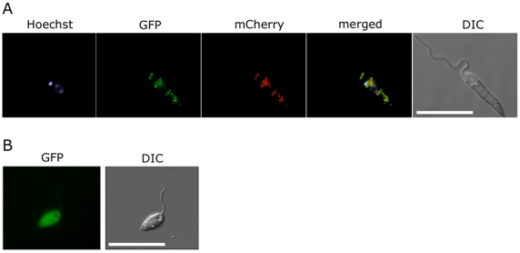
Intracellular parasites, such as Leishmania spp, must acquire suitable carbon sources from the host cell in order to replicate. Here we present evidence that intracellular amastigote stages of Leishmania exploit amino sugars in the phagolysosome of mammalian macrophages as a source of carbon and energy. L. major parasites are capable of using N-acetylglucosamine and glucosamine as primarily carbon sources and contain key enzymes required for conversion of these sugars to fructose-6-phosphate. The last step in this pathway is catalyzed by glucosamine-6-phosphate deaminase (GND), which was targeted to glycosomes via a canonical C-terminal targeting signal when expressed as a GFP fusion protein. Mutant parasites lacking GND were unable to grow in medium containing amino sugars as sole carbohydrate source and rapidly lost viability, concomitant with the hyper-accumulation of hexosamine-phosphates. Expression of native GND, but not a cytosolic form of GND, in Δgnd parasites restored hexosamine-dependent growth, indicating that toxicity is due to depletion of glycosomal pools of ATP. Non-lethal increases in hexosamine phosphate levels in both Δgnd and wild type parasites was associated with a defect in promastigote metacyclogenesis, suggesting that hexosamine phosphate levels may influence parasite differentiation. Promastigote and amastigote stages of the Δgnd mutant were unable to replicate within macrophages and were either completely cleared or exhibited reduced lesion development in highly susceptible Balb/c mice. Our results suggest that hexosamines are a major class of sugars in the macrophage phagolysosome and that catabolism of scavenged amino sugars is required to sustain essential metabolic pathways and prevent hexosamine toxicity.


Conflict of interest statement

The authors have declared that no competing interests exist.

Figures

Figure 1
Figure 1. Hexosamine metabolism in Leishmania.
Exogenous hexosamines (GlcN/GlcNAc) are phosphorylated in the glycosome by a hexose kinase (HK) and either transported to the cytosol for conversion to UDP-GlcNAc or catabolized to Fru6P by the activities of N-acetylglucosamine 6-phosphate deacetylase (NAGD) and glucosamine 6-phosphate deaminase (GND). GND generates fructose-6-phosphate (Fru6P), a key intermediate in carbon metabolism and several pathways of glycoconjugate biosynthesis. The de novo synthesis of GlcNAc6P is catalyzed by the cytoplasmic enzymes, glutamine:Fru6P aminotransferase (GFAT) and glucosamine 6-phosphate acetylase (GNAT).
Figure 2
Figure 2. GND is required for growth on GlcN or GlcNAc.
(A) Wild type (WT), Δgnd and complemented strains (Δgnd +GND, Δgnd +GNDΔSKL were suspended in CDM with or without Glc, GlcN or GlcNAc (13 mM). The optical density of cultures (OD600) at day 4 are shown. (B) L. major wild type, Δgnd, Δgnd +GND and Δgnd +GNDΔSKL promastigotes were lysed and GND activity determined by measuring production of hexose-6-phosphates from GlcN6P. (C,D) Wild type, Δgnd and complemented strains were labeled with 3H-GlcN and incorporation of 3H-label into (C) lipids and (D) the carbohydrate reserve polymer mannogen, assessed by HPTLC and fluorography. Major lipid species comprise PtdEtn, PtdCho, PtdIno, inositolphosphoceramide (IPC), PtdInoP and GPI species (asterix); Mannose (M) and mannogen oligomers (Mn).
Figure 3
Figure 3. Glycosomal targeting of GND.
(A) L. major promastigotes were transfected with plasmid encoding GFP-GND and mCherry-FBP (glycosomal marker) protein and the two proteins localized by fluorescence microscopy and Differential interference contrast (DIC) microscopy after labeling with Hoechst dye (nuclear and mtDNA). (B) Localization of the GFP-GND fusion protein lacking the canonical C-terminal tripeptide sequence SKL. Scale bar  = 10 µm.
Figure 4
Figure 4. Exogenous hexosamines become toxic in the absence of GND.
(A) Wild type, Δgnd and Δgnd complemented with full length GND or truncated GND (GNDΔSKL) were cultivated in M199 medium containing either Glc, GlcN, GlcNAc or no hexoses. Parasite morphology was monitored by DIC microscopy after 24 hr. (B) Wild type and Δgnd promastigotes were cultivated in M199 medium with or without indicated sugars for 24 hr. Parasite survival was determined by suspending parasites in complete medium containing glucose and measuring OD600 at day 2. Survival is expressed relative to wild type parasites grown in glucose-supplemented media from three independent experiments. Error bar  =  SD. Glycerol is abbreviated as Gro.
Figure 5
Figure 5. Accumulation of hexosamine phosphates in GlcNAc-fed Δgnd parasites.
(A) Concentration of intracellular sugar-phosphates in WT and Δgnd parasites after cultivation (4hr) in the presence of Glc or GlcNAc. (B) L. major wild type and Δgnd promastigotes were suspended in hexose-free CDM supplemented with or without 13 mM GlcNAc. After 4 hr, the medium was supplemented with 13 mM [U-13C]-Glc and parasites harvested at the indicated time points. The rate of synthesis of Glc6P (indicated by percent Glc6P labeled with 13C), an indicator of glycolytic flux, was determined by GC-MS.
Figure 6
Figure 6. Non-lethal accumulation of hexosamine phosphates leads to a defect in metacyclogenesis.
(A) WT, Δgnd and Δgnd+GND promastigotes were cultivated in SDM containing 10% FCS and differentiation to metacyclic stages determined after agglutination with the peanut agglutinin lectin (PNA). Unlike dividing promastigotes, metacyclic promastigotes are not agglutinated by PNA. (B) L. major wild type promastigotes were grown in CDM containing either Glc, GlcNAc or GlcN as the sole carbohydrate and metacyclogenesis assessed by PNA agglutination. Error bars represent SED from three independent experiments.
Figure 7
Figure 7. Attenuated virulence of Δgnd in mice and macrophages.
(A) Wild type, Δgnd and complemented Δgnd promastigotes were used to infect BALB/c mice intradermally and lesion formation was scored over time (error bar  =  SEM, n = 5). * p<0.01 (Student t-test). (B) Lesion scores in mice infected with wild type, Δgnd, and complemented Δgnd promastigotes 20 weeks post-infection. The line represents the average of three independent experiments of 15 mice in total. (C) Lesion burden was determined by the limiting dilution assay from draining lymph nodes (parasite numbers are based on 1 million lymph cells). (D) Lesion-derived amastigotes were used to re-infect naïve BALB/c mice and lesions were scored as in (A). RAW 264.7 macrophages were infected with promastigotes of L. major wild type, Δgnd, Δgnd +GND and Δgnd +GNDΔSKL and (E) percent infected macrophages and (F) intracellular parasite numbers were determined by microscopy at day 1, 4 and 6 p.i. Error bars represent SED from three independent experiments. * p<0.05 (Student t-test).
Figure 8
Figure 8. Survival and growth of Δgnd amastigotes in ex vivo macrophages.
BALB/c mouse peritoneal macrophages were infected with lesion-derived amastigotes of L. major wild type, Δgnd, complemented Δgnd (10∶1 ratio parasites/macrophage). Infected macrophages and intracellular parasite growth (as percentage of number of parasites/100 macrophages compared to day 1) were determined by microscopy at day 4 p.i. (Error bars: SED, n = 3).

Similar articles

Cited by

References

    1. Isberg RR, O'Connor TJ, Heidtman M. The Legionella pneumophila replication vacuole: making a cosy niche inside host cells. Nat Rev Microbiol. 2009;7:13–24. - PMC - PubMed
    1. Swanson MS, Fernandez-Moreira E. A microbial strategy to multiply in macrophages: the pregnant pause. Traffic. 2002;3:170–177. - PubMed
    1. Schaible UE, Kaufmann SH. A nutritive view on the host-pathogen interplay. Trends Microbiol. 2005;13:373–380. - PubMed
    1. Appelberg R. Macrophage nutriprive antimicrobial mechanisms. J Leukoc Biol. 2006;79:1117–1128. - PubMed
    1. Naderer T, McConville MJ. The Leishmania-macrophage interaction: a metabolic perspective. Cell Microbiol. 2008;10:301–308. - PubMed

Publication types