. 2010 Dec 23;6(12):e1001249.
doi: 10.1371/journal.ppat.1001249.

Molecular architectures of trimeric SIV and HIV-1 envelope glycoproteins on intact viruses: strain-dependent variation in quaternary structure

Affiliations

Molecular architectures of trimeric SIV and HIV-1 envelope glycoproteins on intact viruses: strain-dependent variation in quaternary structure

Tommi A White et al. PLoS Pathog. .

Abstract

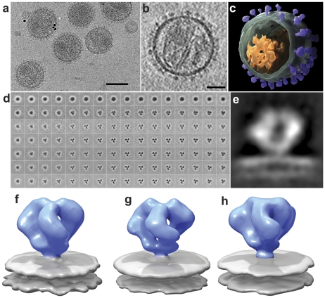
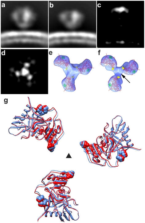
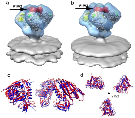
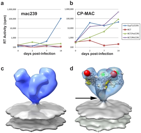
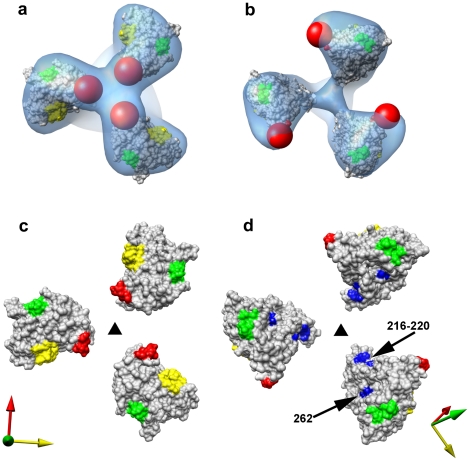
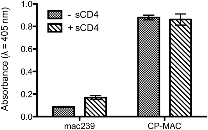
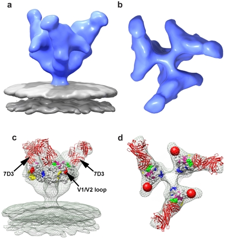
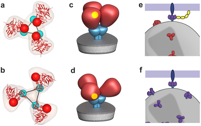
The initial step in target cell infection by human, and the closely related simian immunodeficiency viruses (HIV and SIV, respectively) occurs with the binding of trimeric envelope glycoproteins (Env), composed of heterodimers of the viral transmembrane glycoprotein (gp41) and surface glycoprotein (gp120) to target T-cells. Knowledge of the molecular structure of trimeric Env on intact viruses is important both for understanding the molecular mechanisms underlying virus-cell interactions and for the design of effective immunogen-based vaccines to combat HIV/AIDS. Previous analyses of intact HIV-1 BaL virions have already resulted in structures of trimeric Env in unliganded and CD4-liganded states at ~20 Å resolution. Here, we show that the molecular architectures of trimeric Env from SIVmneE11S, SIVmac239 and HIV-1 R3A strains are closely comparable to that previously determined for HIV-1 BaL, with the V1 and V2 variable loops located at the apex of the spike, close to the contact zone between virus and cell. The location of the V1/V2 loops in trimeric Env was definitively confirmed by structural analysis of HIV-1 R3A virions engineered to express Env with deletion of these loops. Strikingly, in SIV CP-MAC, a CD4-independent strain, trimeric Env is in a constitutively "open" conformation with gp120 trimers splayed out in a conformation similar to that seen for HIV-1 BaL Env when it is complexed with sCD4 and the CD4i antibody 17b. Our findings suggest a structural explanation for the molecular mechanism of CD4-independent viral entry and further establish that cryo-electron tomography can be used to discover distinct, functionally relevant quaternary structures of Env displayed on intact viruses.


Conflict of interest statement

The authors have declared that no competing interests exist.

Figures

Figure 1
Figure 1. Cryo-electron tomography and molecular architecture of trimeric SIV Env.
(a) Low-dose (12 electrons/Å2) projection image recorded from purified SIVmneE11S virions plunge-frozen directly in physiological buffer. The dark spots are 10 nm-sized gold particles that serve as fiducial markers for tilt series alignment. (b) Slice through reconstructed cryo-electron tomograms of a single SIVmneE11S virus. The image was obtained by averaging 6 slices from a 4×4 binned tomogram and is ∼10 nm thick. The viral core and surface spikes can be clearly visualized in the image. (c) 3D rendering of the virion shown in (b), segmented to highlight the viral membrane (green), core (orange) and abundant surface spikes (purple). (d) Results from classification and 3D averaging of ∼4000 3D volumes of SIVmneE11S showing improvement from the first round (second row), to the final round (bottom row) of image classification (see Figure S2 for expanded version of this figure panel). Early stages of classification clearly show classes with inherent 3-fold symmetry, and typically at the fourth iteration, 3-fold symmetry was imposed. At the end of each iteration, the classes that showed the most clearly delineated features in all regions of the spike were selected and combined for use as a reference for the next round. The progression of images from left to right represent successive slices (4.1 Å thickness) from bottom to top of Env through the density map of averaged classes that was used as a reference for the next iteration. The subset of slices shown spans a region of ∼65 Å between the gp120/gp41 interface to the apex of gp120. (e) 2D density profile from a slice through the density map for SIVmneE11S derived from classification and 3D averaging of ∼4000 spikes. (f-h) Perspective views of density maps for SIVmneE11S (f), SIVmac239 (g) and HIV-1 BaL (h) Env shown as isosurface representations with envelope glycoprotein ectodomain shown in blue and viral membrane shown in gray. Scale bars are 100 nm in panel (a) and 35 nm in panel (b).
Figure 2
Figure 2. Structural analysis of full-length and V1/V2 loop-deleted HIV-1 R3A trimeric Env.
(a, b) Side view of raw density projections for full-length trimeric HIV-1 R3A Env (a) and the V1/V2-loop deleted HIV-1 R3A mutant (b) showing the loss of density at the apex in the mutant. This is confirmed by location of the peak in the difference density projections between full-length and loop deleted Env (c, d), shown both as a side view (c) and as a top view (d). (e, f) Top views of fitted density maps rendered as isosurfaces for wild-type and V1/V2 loop-deleted mutants, respectively, of the HIV-1 R3A strain. The gp120 coordinates (1GC1) are shown as red ribbons and were fitted using automated procedures into the density maps. The black arrow in (f) points to the location of the missing density in the V1/V2 loop-deleted variant, and residues in stump of the V1/V2 and V3 loops in the coordinates are highlighted in yellow and in green spheres, respectively. (g) Superposition of fitted coordinates for the gp120 trimer in the full-length and V1/V2 loop-deleted viruses with fits to the wild-type map in red and the mutant in blue, with the black triangle representing the 3-fold symmetry axis.
Figure 3
Figure 3. Molecular architecture of trimeric SIV Env.
(a, b) Perspective views of gp120 monomer coordinates (1GC1) fit into the Env density maps obtained from SIVmneE11S (a) and SIVmac239 (b) using automated procedures. Maps are rendered as transparent isosurfaces with the grey space-filling model of gp120 colored green at base of the V3 loop with the CD4-binding site shown in yellow. The red spheres indicate location of the ∼90 V1/V2 residues of the loop (black arrows) missing in the gp120 coordinates. (c, d) Superposition of the fitted gp120 coordinates for SIVmneE11S (blue ribbons) and SIVmac239 (red ribbons) shown in front (c) view and rotated 90° around the x-axis to display the top (d) view, with the black triangle representing the 3-fold symmetry axis.
Figure 4
Figure 4. Molecular architecture of constitutively open trimeric SIV Env from the CD4-independent CP-MAC strain.
(a,b) Demonstration of CD4-independent viral entry by SIV CP-MAC virus based on infectivity as determined on CD4+ (SupT1) or CD4 (BC7) cells bearing human or rhesus CCR5 for SIVmac239 (a) or SIV CP-MAC (b). Infectivity was determined by reverse transcriptase assays of culture supernatant and confirmed by immunofluorescence assays of cells for SIV-p27gag (Figure S8). (c) Perspective view of density map of Env at ∼20 Å resolution for SIV CP-MAC shown as an isosurface representation. (d) Automated fit of HIV-1 gp120 monomer coordinates (1GC1) into the density map with the CD4 binding site and the base of the V3 loop highlighted in yellow and green, respectively. Absent in the gp120 coordinates are ∼90 residues in the V1/V2 loops, and ∼100 residues in the N- and C-termini, close to the gp120/gp41 interface. The missing V1/V2 loop is represented by the red sphere and the location of the gp120/gp41 interface is indicated by the black arrow.
Figure 5
Figure 5. Closed and open states of trimeric SIV Env.
(a, b) Top views of the density maps of (a) closed (SIVmneE11S) and (b) open (SIV CP-MAC) states fitted with gp120 coordinates. The missing V1/V2 loop is represented by the red sphere to highlight the dramatic difference in location of the loop between the two states. (c, d) Top view of the molecular surfaces of gp120 trimers in (c) closed (SIVmneE11S) and (d) open (SIV CP-MAC) states, highlighting selected residues (216–220, 262, shown in blue) that are conserved across most SIV and HIV-1 Env sequences, which are buried in the closed state, but exposed in the open state. Residues in the CD4 binding site and in the stem of the V1/V2 and V3 loops are shown in yellow, red and green, respectively. The coordinate system in each panel provides an aid to visualizing the rotation of each gp120 monomer in the transition from the closed to open state. Filled dark triangle indicates location of the 3-fold symmetry axis.
Figure 6
Figure 6. ELISA analysis comparing the binding of 7D3 to either SIVmac239 or SIV CP-MAC viruses.
The binding analyses, carried out under the same conditions as the tomographic experiments, show that in contrast to SIVmac239, SIV CP-MAC binds efficiently to 7D3 in the absence and presence of added sCD4-183.
Figure 7
Figure 7. Molecular architecture of co-receptor-binding site antibody (7D3) complexes on SIV CP-MAC Env and fitted gp120 coordinates.
(a, b) Perspective (a) and top (b) views of 7D3-bound SIV CP-MAC Env rendered as isosurfaces. (c, d) Fit of gp120 coordinates shown as isosurfaces to the density maps (green mesh) for 7D3-bound SIV CP-MAC Env rendering from perspective (c) and top views (d). The coordinates for 17b Fab were utilized for representing 7D3 (shown in red ribbons) and fit into the difference density calculated between the antibody-bound and corresponding unliganded SIV CP-MAC Env maps. Residues in the CD4 binding site and in the stem of the V1/V2 and V3 loops are shown in yellow, red and green, respectively, with proposed coreceptor-binding site (CoRbs) residues shown in magenta.
Figure 8
Figure 8. Distinct quaternary conformational states of trimeric Env.
(a,b) Top views of fitted density maps for experimentally derived maps of trimeric Env representing the “closed” states of SIVmneE11S (a), and the “open” state of SIV CP-MAC (b). Coordinates for the gp120 core are shown in red ribbons, density maps shown in transparent brown. The likely locations of the gp120/gp41 interface are indicated by linked cyan spheres, whereas V1/V2 loops are indicated by red spheres. (c,d) Schematic view of trimeric envelope glycoprotein spikes showing the closed (c) and open (d) states, with gp120, gp41, and CD4-binding site in red, cyan, and yellow, respectively. (e,f) Schematic comparison of the mechanism of cell-virus contact in CD4-dependent vs. CD4-independent viral entry. For CD4-dependent entry (e), the transition of the spike from the closed (red) to open (purple) state occurs in the presence of CD4 (yellow) and co-receptor (blue) binding, while in CD4-independent entry (f), trimeric Env is already in a conformation capable of binding to co-receptor.

References

    1. Wyatt R, Sodroski J. The HIV-1 envelope glycoproteins: fusogens, antigens, and immunogens. Science. 1998;280:1884–1888. - PubMed
    1. Dalgleish AG, Beverley PC, Clapham PR, Crawford DH, Greaves MF, et al. The CD4 (T4) antigen is an essential component of the receptor for the AIDS retrovirus. Nature. 1984;312:763–767. - PubMed
    1. Burton DR, Desrosiers RC, Doms RW, Koff WC, Kwong PD, et al. HIV vaccine design and the neutralizing antibody problem. Nat Immunol. 2004;5:233–236. - PubMed
    1. Sato S, Johnson W. Antibody-mediated neutralization and Simian immunodeficiency virus models of HIV/AIDS. Current HIV Research. 2007;5:594–607. - PubMed
    1. Chen B, Vogan EM, Gong H, Skehel JJ, Wiley DC, et al. Structure of an unliganded simian immunodeficiency virus gp120 core. Nature. 2005;433:834–841. - PubMed

Publication types