. 2010 Dec 20;5(12):e15636.
doi: 10.1371/journal.pone.0015636.

Praf2 is a novel Bcl-xL/Bcl-2 interacting protein with the ability to modulate survival of cancer cells

Affiliations

Praf2 is a novel Bcl-xL/Bcl-2 interacting protein with the ability to modulate survival of cancer cells

Maria Teresa Vento et al. PLoS One. .

Abstract

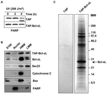
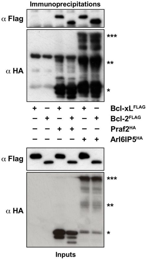
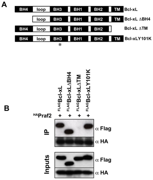
Increased expression of Bcl-xL in cancer has been shown to confer resistance to a broad range of apoptotic stimuli and to modulate a number of other aspects of cellular physiology, including energy metabolism, cell cycle, autophagy, mitochondrial fission/fusion and cellular adhesion. However, only few of these activities have a mechanistic explanation. Here we used Tandem Affinity purification to identify novel Bcl-xL interacting proteins that could explain the pleiotropic effects of Bcl-xL overexpression. Among the several proteins co-purifying with Bcl-xL, we focused on Praf2, a protein with a predicted role in trafficking. The interaction of Praf2 with Bcl-xL was found to be dependent on the transmembrane domain of Bcl-xL. We found that Bcl-2 also interacts with Praf2 and that Bcl-xL and Bcl-2 can interact also with Arl6IP5, an homologue of Praf2. Interestingly, overexpression of Praf2 results in the translocation of Bax to mitochondria and the induction of apoptotic cell death. Praf2 dependent cell death is prevented by the co-transfection of Bcl-xL but not by its transmembrane domain deleted mutant. Accordingly, knock-down of Praf2 increases clonogenicity of U2OS cells following etoposide treatment by reducing cell death. In conclusion a screen for Bcl-xL-interacting membrane proteins let us identify a novel proapoptotic protein whose activity is strongly counteracted exclusively by membrane targeted Bcl-xL.


Conflict of interest statement

Competing Interests: The authors have declared that no competing interests exist.

Figures

Figure 1
Figure 1. Bcl-xL is present in a wide range of molecular weights in fractionated U2OS cell extracts.
Sucrose gradient fractionation of U2OS total cell extracts. U2OS (60×106 cells) were lysed in CHAPS extraction buffer and cleared by centrifugation. Extracts were fractionated using a 10–40% sucrose gradient and equal volumes of the fractions were analysed by immunoblot for the presence of Bcl-xL, Bax, Rab4 and Cytochrome c.
Figure 2
Figure 2. TAP-Bcl-xL is able to protect from apoptosis, localise correctly and co-purify with several cellular proteins.
(A) HeLa cells stably expressing TAP-tagged Bcl-xL are more resistant than control to UV-induced apoptosis. Analysis of PARP cleavage in HeLa cells expressing TAP alone or TAP-Bcl-xL and irradiated or not with UV (200 J/m2). Cells were collected 2 and 4 hours after irradiation and analysed by immunoblot for the appearance of the cleaved product of PARP. (B) TAP-tagged Bcl-xL localises to the same intracellular compartment of endogenous Bcl-xL. HeLa cells expressing TAP-Bcl-xL were fractionated by differential centrifugation to obtain: lane 1, cytosolic proteins (S100); lane 2, nuclear proteins (Nuclei); lane 3, the heavy membrane fraction (HMM); lane 4, the light membrane fraction (LMM). 20 µg of each fraction were analysed by immunoblot for the presence of TAP-BclxL (using secondary antibody only) and endogenous Bcl-xL. The quality of the fractionation was determined using the antibodies against Sec23 (LMM), Cytochrome c (HMM), Bax (S100 and HMM) and PARP (Nuclei). (C) HeLa cells stably transfected with TAP alone or TAP-Bcl-xL were subjected to Tandem Affinity Purification. The figure shows a representative Coomassie stained SDS-PAGE performed under reducing conditions used to resolve eluates from purifications. Purifications were performed using membrane CHAPS extracts from HeLa expressing TAP alone (lane 1) or HeLa expressing TAP-Bcl-xL (lane 2).
Figure 3
Figure 3. Bcl-xL and Praf2 interact by co-immunoprecipitation.
(A) HEK 293T cells were either transfected with HA-tagged Praf2 or FLAG-tagged BclxL or co-transfected with both expression vectors. Samples were subjected to immunoprecipitation, using anti HA-conjugated agarose and both total lysates (inputs) and agarose beads eluates (IP) were analysed by immunoblot using anti HA and anti FLAG monoclonal antibodies. (B) U2OS cells were transfected with HA-tagged Praf2 or the empty vector (Vec) and cell lysates were subjected to immunoprecipitation, using HA-conjugated agarose. Both lysates and agarose beads were analysed by immunoblot using anti HA and anti Bcl-xL antibodies.
Figure 4
Figure 4. Praf2 and Arl6IP5 interact also with Bcl-2.
HEK 293 cells were either transfected with FLAG-tagged BclxL or Bcl-2 alone (lane 1 and 2), or co-transfected with HA-tagged Praf2 or HA-tagged Arl6IP5 (lanes 3 to 6). Samples were subjected to immunoprecipitation, using HA-conjugated agarose and both lysates and agarose beads were analysed by immunoblot using anti HA and anti FLAG antibodies. Arl6IP5 monomer: *; Arl6IP5 dimer: **; Arl6IP5 multimer: ***.
Figure 5
Figure 5. Deletion of Bcl-xL's transmembrane domain prevents its interaction with Praf2.
(A) Schematic representation of the Bcl-xL expression constructs used in this study. Bcl-xL: full length Bcl-xL; Bcl-xLΔBH4: Bcl-xL lacking the first 24 aminoacids; Bcl-xLΔTM: Bcl-xL lacking the last 22 aminoacids; Bcl-xLY101K: Bcl-xL with a substitution of Tyrosine 101 with Lysine. (B) HEK 293 cells were co-transfected with HA-tagged Praf2 and the BclxL constructs described in (A) carrying a FLAG-tag at the N-terminus. Samples were subjected to immunoprecipitation, using HA-conjugated agarose and both lysates and agarose beads were analysed by immunoblot using anti HA and anti FLAG antibodies.
Figure 6
Figure 6. Expression of Praf2 induces apoptotic cell death.
(A) FACS analysis of HeLa cells transfected with the indicated expression vectors and stained with Propidium Iodide (PI). Percentages of PI positive cells are indicated on the top-left of each graph. (B) Aliquots of the HeLa cells transfected as in (A) were analysed by immunoblot for the appearance of the cleaved form of PARP. Expression levels of Praf2, Bcl-xL and Bcl-xLΔTM are also indicated. (C) Analysis of GFP-Bax localisation in U2OS cells transfected with empty vector (vec) or HA-tagged Praf2. The photographs are representative of the two main GFP-Bax localisations observed in the samples: diffuse cytosolic and aggregated. Above the photographs are indicated the % of GFP positive cells with an aggregated localisation after transfecting cells with empty vector or HA-tagged Praf2 in two independent experiments.
Figure 7
Figure 7. Praf2 knock down reduces apoptosis and increases clonogenicity of U2OS cells treated with etoposide.
(A) Analysis by immunoblot of the efficacy of Praf2 knock down following transfection of U2OS cells with two different siRNAs targeting the Praf2 mRNA. (B) U2OS cells transfected with control (Ctrl) or Praf2 targeting siRNAs (siRNA5 and siRNA6) after 48 hours were treated with 50 µM etoposide for 24 hours. Cellular caspase 3 and 7 activities were measured using the Caspase-Glo 3/7 luminometric assay. The graph show the percentage of caspase 3 and 7 activation in samples treated with the Praf2 targeting siRNAs compared with the activity present in cells transfected with the control siRNA in 3 independent experiments. (C) Clonogenicity of U2OS cells transfected with control or Praf2 targeting siRNAs and treated or not with etoposide. 48 hours after transfection of U2OS cells with control siRNA or Praf2 siRNA6, cells were treated with 50 µM etoposide for 3 hours and than shifted again to normal medium for 2 weeks. Colonies were colored using crystal violet. The graph represents the percentage of colonies grown after etoposide treatment respect to the untreated control.

References

    1. Iaccarino I, Hancock D, Evan G, Downward J. c-Myc induces cytochrome c release in Rat1 fibroblasts by increasing outer mitochondrial membrane permeability in a Bid-dependent manner. Cell Death Differ. 2003;10:599–608. - PubMed
    1. Dong Z, Wang J. Hypoxia selection of death-resistant cells. A role for Bcl-X(L). J Biol Chem. 2004;279:9215–9221. - PubMed
    1. Kauffmann-Zeh A, Rodriguez-Viciana P, Ulrich E, Gilbert C, Coffer P, et al. Suppression of c-Myc-induced apoptosis by Ras signalling through PI(3)K and PKB. Nature. 1997;385:544–548. - PubMed
    1. Amundson SA, Myers TG, Scudiero D, Kitada S, Reed JC, et al. An Informatics Approach Identifying Markers of Chemosensitivity in Human Cancer Cell Lines. Cancer Res. 2000;60:6101–6110. - PubMed
    1. Youle RJ, Strasser A. The BCL-2 protein family: opposing activities that mediate cell death. Nat Rev Mol Cell Biol. 2008;9:47–59. - PubMed

Publication types