. 2010 Dec 21;8(12):e1000563.
doi: 10.1371/journal.pbio.1000563.

HER2 phosphorylation is maintained by a PKB negative feedback loop in response to anti-HER2 herceptin in breast cancer

Affiliations

HER2 phosphorylation is maintained by a PKB negative feedback loop in response to anti-HER2 herceptin in breast cancer

Merel Gijsen et al. PLoS Biol. .

Erratum in

Abstract

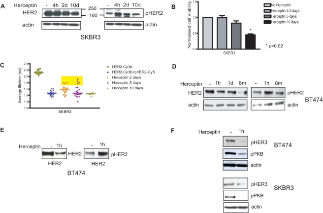
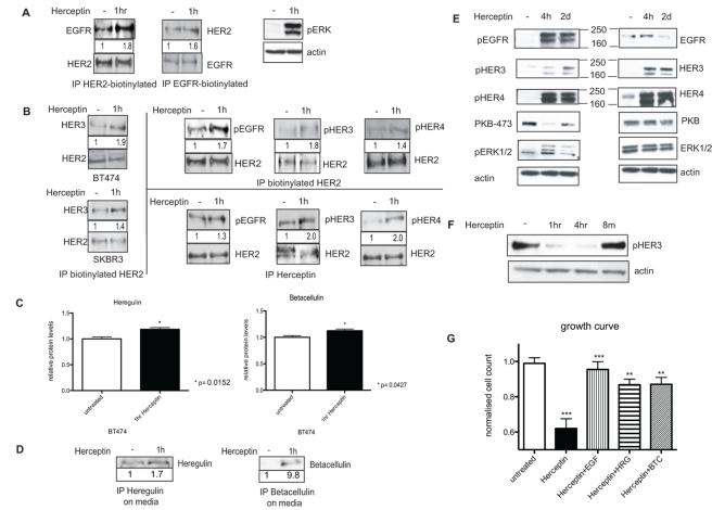
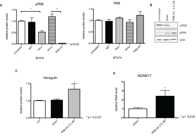
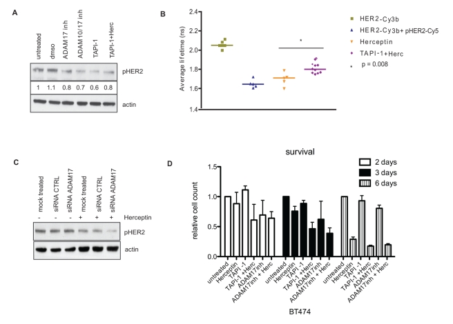
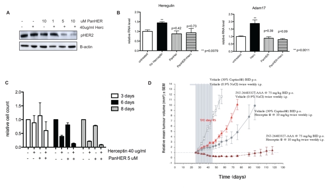
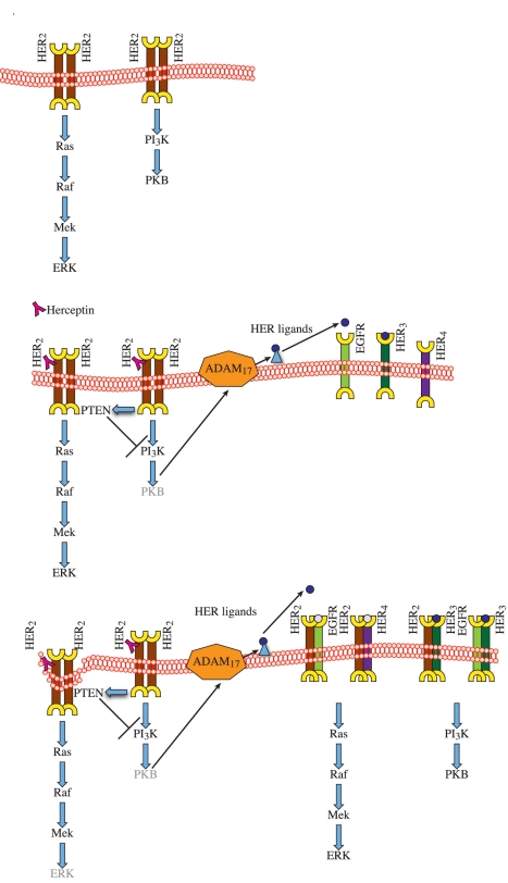
Herceptin (trastuzumab) is used in patients with breast cancer who have HER2 (ErbB2)-positive tumours. However, its mechanisms of action and how acquired resistance to Herceptin occurs are still poorly understood. It was previously thought that the anti-HER2 monoclonal antibody Herceptin inhibits HER2 signalling, but recent studies have shown that Herceptin does not decrease HER2 phosphorylation. Its failure to abolish HER2 phosphorylation may be a key to why acquired resistance inevitably occurs for all responders if Herceptin is given as monotherapy. To date, no studies have explained why Herceptin does not abolish HER2 phosphorylation. The objective of this study was to investigate why Herceptin did not decrease HER2 phosphorylation despite being an anti-HER2 monoclonal antibody. We also investigated the effects of acute and chronic Herceptin treatment on HER3 and PKB phosphorylation in HER2-positive breast cancer cells. Using both Förster resonance energy transfer (FRET) methodology and conventional Western blot, we have found the molecular mechanisms whereby Herceptin fails to abolish HER2 phosphorylation. HER2 phosphorylation is maintained by ligand-mediated activation of EGFR, HER3, and HER4 receptors, resulting in their dimerisation with HER2. The release of HER ligands was mediated by ADAM17 through a PKB negative feedback loop. The feedback loop was activated because of the inhibition of PKB by Herceptin treatment since up-regulation of HER ligands and ADAM17 also occurred when PKB phosphorylation was inhibited by a PKB inhibitor (Akt inhibitor VIII, Akti-1/2). The combination of Herceptin with ADAM17 inhibitors or the panHER inhibitor JNJ-26483327 was able to abrogate the feedback loop and decrease HER2 phosphorylation. Furthermore, the combination of Herceptin with JNJ-26483327 was synergistic in tumour inhibition in a BT474 xenograft model. We have determined that a PKB negative feedback loop links ADAM17 and HER ligands in maintaining HER2 phosphorylation during Herceptin treatment. The activation of other HER receptors via ADAM17 may mediate acquired resistance to Herceptin in HER2-overexpressing breast cancer. This finding offers treatment opportunities for overcoming resistance in these patients. We propose that Herceptin should be combined with a panHER inhibitor or an ADAM inhibitor to overcome the acquired drug resistance for patients with HER2-positive breast cancer. Our results may also have implications for resistance to other therapies targeting HER receptors.


Conflict of interest statement

Tim Perera and Peter King are employees and small share-holders of Johnson & Johnson Pharmaceutical Research & Development. This Competing Interest does not alter our adherence to all the PLoS Biology policies on sharing data and materials, or to the Creative Commons Attribution License in the event of publication.

Figures

Figure 1
Figure 1. Herceptin down-regulates HER2 receptors and decreases HER3 phosphorylation, but it does not decrease HER2 phosphorylation.
(A) SKBR3 cells were lysed for Western blot analysis after pre-treatment with 40 µg/ml Herceptin for a period of 4 h, 2 d, or 10 d. Equal amounts of protein were loaded in each lane, and two SDS-PAGE gels were run. Membranes were blotted with appropriate antibodies for total and phosphorylated HER2 and actin. (B) SKBR3 cells were grown in 24-well plates and left to grow for at least 24 h before being treated with 40 µg/ml Herceptin for different durations, as illustrated. The viable cells were counted in a cell viability analyzer using trypan blue to stain the dead cells. (C) SKBR3 cells were incubated with either donor alone (HER2-Cy3b) or donor and acceptor (HER2-Cy3b+pHER2-Cy5) to assess HER2 phosphorylation by FRET after pre-treatment with 40 µg/ml Herceptin for different durations, as illustrated. (D) BT474 cells were lysed for Western blot analysis after pre-treatment with 40 µg/ml Herceptin for a period of 1 h, 1 d, or 8 mo with replacement every week. Equal amounts of protein were loaded in each lane. Membranes were blotted with antibodies for total and phosphorylated HER2 and actin. (E) BT474 cells were immunoprecipitated with an antibody for cytoplasmic anti-HER2 (1∶100). Following the immunoprecipitation the cell lysate was loaded on an SDS gel. The membrane was probed with antibodies for total or phosphorylated HER2. (F) BT474 and SKBR3 cells were lysed after pre-treatment with 40 µg/ml Herceptin for 1 h. Equal amounts were loaded on an SDS gel, and membranes were probed for phosphorylated HER3, phosphorylated PKB, and actin.
Figure 2
Figure 2. Herceptin induces the activation of HER receptors and their dimerisation with HER2 as a result of an up-regulation and the release of HER ligands.
(A) In the left two panels, BT474 cells were immunoprecipitated (IP) with either intracellular anti-EGFR or anti-HER2 antibody after 1 h of 40 µg/ml Herceptin treatment. Following the immunoprecipitation, the cell lysate was loaded unto an SDS gel, and a Western blot analysis was performed. The membrane was probed with either anti-EGFR or anti-HER2 antibody, as illustrated. In the right panel, BT474 cells treated for 1 h with 40 µg/ml Herceptin were lysed, and equal amounts were loaded on a gel. Membrane was probed for phosphorylated ERK and actin. (B) In the left upper and lower panels, BT474 and SKBR3 cells were immunoprecipitated with biotinylated anti-HER2 antibody after the cells were treated for 1 h with 40 µg/ml Herceptin. Following the immunoprecipitation, equal amounts of the cell lysates were loaded unto an SDS gel, a Western blot analysis was performed, and the membrane was probed with anti-HER3 antibody. In the top right three panels, BT474 cells were immunoprecipitated with biotinylated anti-HER2 antibody after 1 h of Herceptin treatment. Western blot analysis was done using antibodies that recognised pEGFR, pHER3, or pHER4. In the lower right three panels, BT474 cells were treated with 40 µg/ml Herceptin for 1 h before the lysate was immunoprecipitated using the anti-HER2 antibody Herceptin. After immunoprecipitation, lysate was loaded onto an SDS gel. A Western blot analysis was performed, and the membrane was probed with anti-pEGFR, anti-pHER3, or anti-pHER4 antibodies. (C) BT474 cells were treated for 1 h with 40 µg/ml Herceptin, and the cells were lysed using Hepes buffer and homogenised before analysing the levels of heregulin and betacellulin by ELISA. (D) In the left panel, serum-free medium of untreated and Herceptin-treated SKBR3 cells was immunoprecipitated for heregulin. A Western blot analysis was then performed, and the membrane was probed for heregulin. In the right panel the same technique was used to look at betacellulin levels in the medium of SKBR3 cells. (E) SKBR3 cells were lysed for Western blot analysis after pre-treatment with 40 µg/ml Herceptin. An equal amount of protein was loaded in each lane. Four parallel SDS-PAGE gels were run, and the membranes were cut in different parts according to molecular weight to analyse the total and phosphorylation levels of HER receptors, PKB, ERK, and actin using appropriate antibodies. (F) BT474 cells were lysed after pre-treatment with 40 µg/ml Herceptin for 1 h, 4 h, or 8 mo. Equal amounts were loaded on an SDS gel, and membranes were probed for phosphorylated HER3 and actin. (G) BT474 cells were treated for 5 d with either 40 µg/ml Herceptin alone or concurrent 40 µg/ml Herceptin treatment with exogenous 100 ng/ml EGF, betacellulin (BTC), or heregulin (HRG) stimulation. Cells were then counted using a cell counter to assess cell viability. For statistical analysis, untreated and Herceptin-treated samples were compared using the Mann-Whitney test. The Herceptin-treated samples were then compared with samples treated with Herceptin and concurrent growth factor stimulation.
Figure 3
Figure 3. Up-regulation of heregulin is mediated by ADAM17, and acute Herceptin treatment induces an increase in mRNA and protein levels of ADAM17.
(A) SKBR3 cells were transfected with siRNA against ADAM17 or a control sequence. The knockdown was validated by Western blot for ADAM17 and actin (left panel). Knockdown cells were then treated with 40 µg/ml Herceptin for 1 h and lysed in Hepes buffer for heregulin detection using ELISA (right panel). (B) SKBR3 cells were treated with 40 µg/ml Herceptin for 1 h, and ADAM17 mRNA levels were studied by QPCR. The Mann-Whitney test was performed to determine statistical significance of the up-regulation of ADAM17 mRNA after Herceptin treatment. (C) BT474 cells (left two panels) and SKBR3 cells (right two panels) were treated with an increasing dose of Herceptin over a period of 1 h. Cell lysate was loaded onto an SDS gel, and a Western blot analysis was performed. The membrane was probed with anti-ADAM17 and anti-pPKB antibodies. Quantification of three separate experiments is depicted in graphs for pPKB and ADAM17; representative blots are shown above the graphs.
Figure 4
Figure 4. Inhibiting PKB phosphorylation by a PKB inhibitor induces up-regulation of heregulin and ADAM17.
(A) BT474 cells were treated for 1 h with 40 µg/ml Herceptin (Herc), 2.5 µM PKB inhibitor (PKB inh), 40 µg/ml IgG control, or DMSO control at 37°C. After 1 h, cells were lysed and analysed for total and phosphorylated PKB levels using MSD multiplex kits. (B) BT474 cell lysates were loaded on an SDS gel, and the membranes were probed for phosphorylated ERK, PKB, and actin. (C) BT474 cells pre-treated with PKB inhibitor were lysed in Hepes buffer and homogenised before analysing protein levels of heregulin using ELISA. (D) BT474 cells were treated with PKB inhibitor for 1 h, and ADAM17 mRNA levels were studied by QPCR. Statistical significance was determined using the Mann-Whitney test.
Figure 5
Figure 5. Combination of Herceptin with ADAM inhibitors decreases HER2 phosphorylation and is additive in cell viability inhibition.
(A) SKBR3 cells were treated with 100 µM ADAM17 inhibitor (inh), 100 µM ADAM10/17 inhibitor, 100 µM ADAM inhibitor TAPI-1, or a combination of TAPI-1 and 40 µg/ml Herceptin (Herc) for 1 h. Cells were then lysed, and equal amounts of protein were loaded on an SDS gel. Membrane was probed for phosphorylated HER2 and actin. (B) FRET experiments to assess HER2 phosphorylation in SKBR3 cells. The cells were treated with 40 µg/ml Herceptin for 1 h with or without TAPI-1. The medians of the lifetimes were compared with the basal condition using the Mann-Whitney test. (C) SKBR3 cells were transfected with siRNA against ADAM17 or a control sequence. Three days after transfection, cells were treated with 40 µg/ml Hercpetin for 1 h. Lysates were loaded on an SDS gel. The membrane was probed for pHER2 and actin. (D) BT474 cells were grown in 24-well plates and left to grow for at least 24 h before being treated for different durations with 40 µg/ml Herceptin, 10 µM TAPI-1, 10 µM ADAM17 inhibitor, or a combination of ADAM inhibitors with Herceptin, as illustrated. The viable cells were counted using a cell counter.
Figure 6
Figure 6. Combination of Herceptin with a panHER inhibitor abrogates the PKB feedback loop and is synergistic in inhibition of xenograft tumour growth.
(A) SKBR3 cells were treated for 1 h with 40 µg/ml Herceptin (Herc) alone, 10 µM panHER inhibitor JNJ-26483327 alone, or Herceptin with 1, 5, or 10 µM JNJ-26483327. Cell lysate was loaded onto an SDS gel, and a Western blot analysis was performed. Membranes were blotted with antibodies recognising pHER2 and β-actin. (B) BT474 cells are treated with 40 µg/ml Herceptin, 5 µM JNJ-26483327, or a combination of both drugs for 1 h. ADAM17 mRNA and heregulin mRNA levels were studied by QPCR. p-values were calculated using the Mann-Whitney test comparing treated samples against untreated samples. (C) BT474 cells were grown in a 24-well plate and left to grow for at least 24 h before being treated for different durations with 40 µg/ml Herceptin, 5 µM JNJ-26483327, or a combination of these drugs, as illustrated. The viable cells were counted using a cell counter. (D) BT474 xenografts were treated with an empty vehicle, JNJ-26483327 (75 mg/kg twice a day orally [BID p.o.]), Herceptin (10 mg/kg twice weekly intraperitonealy [i.p.]), or a combination of JNJ-26483327 with Herceptin treatment for 21 d and were observed for a period of 120 d.
Figure 7
Figure 7. The proposed model of acquired resistance to Herceptin in HER2-overexpressing breast cancer.
In the top diagram, HER2 overexpression (both homodimers and heterodimers) leads to constitutive activation of EGFR and HER2 with MAPK activation . These cells have also been shown to have activated Src with constitutively suppressed PTEN activity and increased PKB activity ,. In the middle diagram, Herceptin increases PTEN membrane localisation and PTEN activation in HER2-overexpressing cells, resulting in suppression of PKB phosphorylation . This inhibition induces the PKB negative feedback loop, triggering the production and release of HER ligands through ADAM17 protease. In the bottom diagram, Herceptin down-regulates HER2 receptors, but the up-regulation of HER ligands causes activation of EGFR, HER3, and HER4 and their dimerisation with HER2, resulting in the maintenance of HER2 phosphorylation, with increased activation of PKB and MAPK pathways.

References

    1. Mendelsohn J, Baselga J. Epidermal growth factor receptor targeting in cancer. Semin Oncol. 2006;33:369–385. - PubMed
    1. Citri A, Yarden Y. EGF-ERBB signalling: towards the systems level. Nat Rev Mol Cell Biol. 2006;7:505–516. - PubMed
    1. Graus-Porta D, Beerli R. R, Daly J. M, Hynes N. E. ErbB-2, the preferred heterodimerization partner of all ErbB receptors, is a mediator of lateral signaling. EMBO J. 1997;16:1647–1655. - PMC - PubMed
    1. Cho H. S, Mason K, Ramyar K. X, Stanley A. M, Gabelli S. B, et al. Structure of the extracellular region of HER2 alone and in complex with the Herceptin Fab. Nature. 2003;421:756–760. - PubMed
    1. Slamon D. J, Clark G. M, Wong S. G, Levin W. J, Ullrich A, et al. Human breast cancer: correlation of relapse and survival with amplification of the HER-2/neu oncogene. Science. 1987;235:177–182. - PubMed

Publication types

MeSH terms