

Localization of AQP5 during development of the mouse submandibular salivary gland
- PMID: 21203896
- PMCID: PMC3063871
- DOI: 10.1007/s10735-010-9308-0
Localization of AQP5 during development of the mouse submandibular salivary gland
Abstract
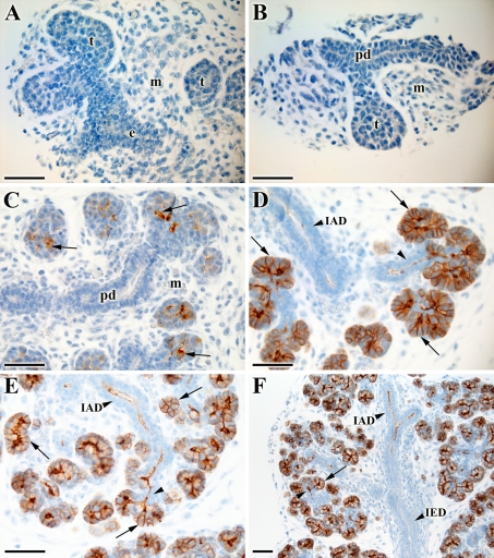
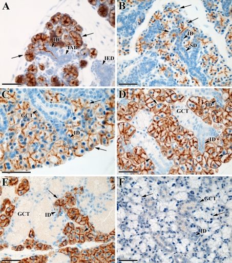
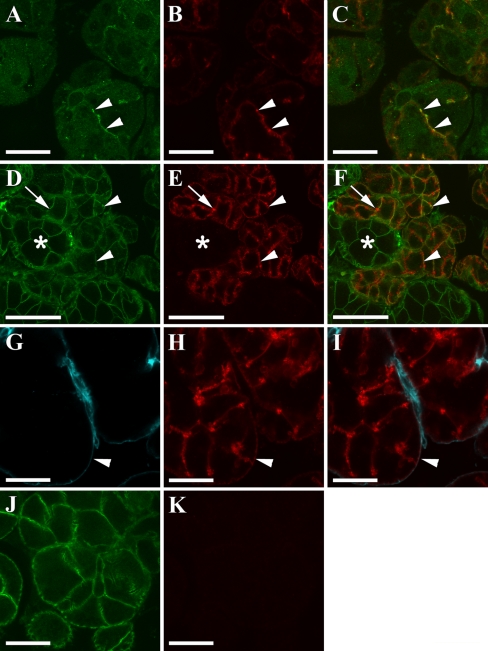
Aquaporin 5 (AQP5) is known to be central for salivary fluid secretion. A study of the temporal-spatial distribution of AQP5 during submandibular gland (SMG) development and in adult tissues might offer further clues to its unknown role during development. In the present work, SMGs from embryonic day (E) 14.5-18.5 and postnatal days (P) 0, 2, 5, 25, and 60 were immunostained for AQP5 and analyzed using light microscopy. Additional confocal and transmission electron microscopy were performed on P60 glands. Our results show that AQP5 expression first occurs in a scattered pattern in the late canalicular stage and becomes more prominent and organized in the terminal tubuli/pro-acinar cells towards birth. Additional apical membrane staining in the entire intralobular duct is found just prior to birth. During postnatal development, AQP5 is expressed in both the luminal and lateral membrane of pro-acinar/acinar cells. AQP5 is also detected in the basal membrane of acinar cells at P25 and P60. In the intercalated ducts at P60, the male glands show apical staining in the entire segment, while only the proximal region is positive in the female glands. These results demonstrate an evolving distribution of AQP5 during pre- and postnatal development in the mouse SMGs.
Figures

Similar articles
-
Aquaporin 5 distribution pattern during development of the mouse sublingual salivary gland.J Mol Histol. 2011 Oct;42(5):401-8. doi: 10.1007/s10735-011-9343-5. Epub 2011 Aug 5. J Mol Histol. 2011. PMID: 21818558
-
Unilateral maxillary molar extraction influences AQP5 expression and distribution in the rat submandibular salivary gland.Arch Oral Biol. 2012 Jul;57(7):877-83. doi: 10.1016/j.archoralbio.2012.02.006. Epub 2012 Mar 14. Arch Oral Biol. 2012. PMID: 22424887
-
Modified aquaporin 5 expression and distribution in submandibular glands from NOD mice displaying autoimmune exocrinopathy.Arthritis Rheum. 2007 Aug;56(8):2566-74. doi: 10.1002/art.22826. Arthritis Rheum. 2007. PMID: 17665453
-
Dynamics of Salivary Gland AQP5 under Normal and Pathologic Conditions.Int J Mol Sci. 2020 Feb 11;21(4):1182. doi: 10.3390/ijms21041182. Int J Mol Sci. 2020. PMID: 32053992 Free PMC article. Review.
-
Physiological role of aquaporin 5 in salivary glands.Pflugers Arch. 2016 Apr;468(4):519-39. doi: 10.1007/s00424-015-1749-6. Epub 2015 Nov 5. Pflugers Arch. 2016. PMID: 26537593 Review.
Cited by
-
A fluid secretion pathway unmasked by acinar-specific Tmem16A gene ablation in the adult mouse salivary gland.Proc Natl Acad Sci U S A. 2015 Feb 17;112(7):2263-8. doi: 10.1073/pnas.1415739112. Epub 2015 Feb 2. Proc Natl Acad Sci U S A. 2015. PMID: 25646474 Free PMC article.
-
Biocompatible tissue scaffold compliance promotes salivary gland morphogenesis and differentiation.Tissue Eng Part A. 2014 Jun;20(11-12):1632-42. doi: 10.1089/ten.TEA.2013.0515. Epub 2014 Feb 27. Tissue Eng Part A. 2014. PMID: 24410370 Free PMC article.
-
Insights into the Function of Aquaporins in Gastrointestinal Fluid Absorption and Secretion in Health and Disease.Cells. 2023 Aug 29;12(17):2170. doi: 10.3390/cells12172170. Cells. 2023. PMID: 37681902 Free PMC article. Review.
-
Organ-specific extracellular matrix directs trans-differentiation of mesenchymal stem cells and formation of salivary gland-like organoids in vivo.Stem Cell Res Ther. 2022 Jul 15;13(1):306. doi: 10.1186/s13287-022-02993-y. Stem Cell Res Ther. 2022. PMID: 35841112 Free PMC article.
-
Neuronal-epithelial cross-talk drives acinar specification via NRG1-ERBB3-mTORC2 signaling.Dev Cell. 2022 Nov 21;57(22):2550-2565.e5. doi: 10.1016/j.devcel.2022.10.011. Dev Cell. 2022. PMID: 36413949 Free PMC article.
References
-
- Abate E, Krane C, Rizvi T, Colbert M, Jiang M, Boivin G, Menon A. Tissue distribution of aquaporin 5 in the developing salivary gland and lung of mouse embryo. J Invest Med. 2003;51:S143.
-
- Akamatsu T, Parvin MN, Murdiastuti K, Kosugi-Tanaka C, Yao CJ, Miki O, Kanamori N, Hosoi K. Expression and localization of aquaporins, members of the water channel family, during development of the rat submandibular gland. Pflugers Arch-Euro J Physiol. 2003;446:641–651. doi: 10.1007/s00424-003-1109-9. - DOI - PubMed
Publication types
MeSH terms
Substances
Grants and funding
LinkOut - more resources
Full Text Sources
Other Literature Sources
Molecular Biology Databases
