Review
. 2011 May;50(1):10-21.
doi: 10.1007/s12026-010-8201-y.

Biology and function of neuroimmune semaphorins 4A and 4D

Affiliations
Review

Biology and function of neuroimmune semaphorins 4A and 4D

EusebiusHenry Nkyimbeng-Takwi et al. Immunol Res. 2011 May.

Abstract

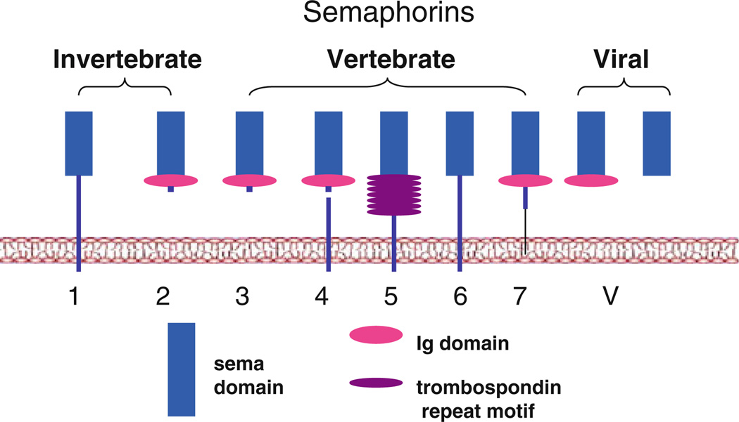
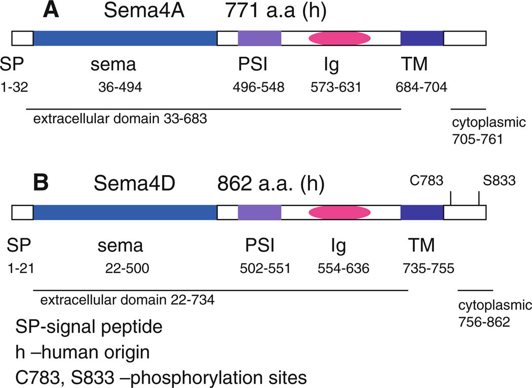
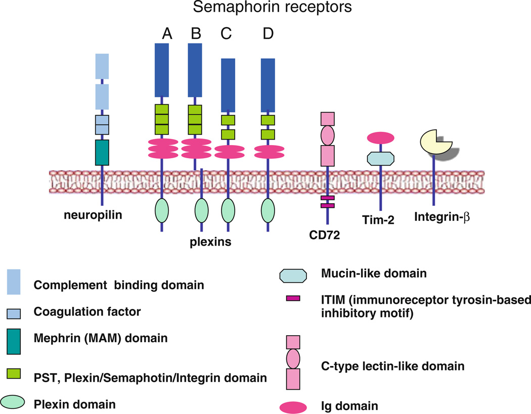
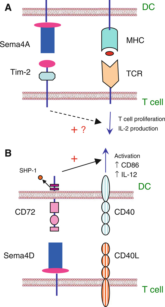
Semaphorins belong to a family of membrane-bound and secreted molecules that regulate the functional activity of axons in the nervous system. Sema4A and Sema4D were the first semaphorins also found to be expressed in immune cells and were, therefore, termed "immune semaphorins". It is known that Sema4A has three functional receptors, namely Plexin D1, Plexin B1, and Tim-2, whereas Sema4D binds to Plexin B1 and CD72. Recent studies suggest that immune semaphorins play critical roles in many physiological and pathological processes and such. In this review, we summarize the current knowledge on the biology of neuroimmune semaphorins and their corresponding receptors, their distribution in organs and tissues, function in the immune response, and critical regulatory roles in various diseases.


Figures

Fig. 1
Fig. 1
Semaphorin family. The members of semaphorin family of secreted and transmembrane proteins have been divided into 8 classes based on origins and structural features. Vertebrates have members in classes 3–7, invertebrates have members in classes 1 and 2, whereas class V semaphorins were found only in viruses. All semaphorins contain a homologous sema sequence of approximately 500 a.a. in N-terminal extracellular domain. They have some distinguishing sequences such as a single C2-class Ig-like domain (classes 3, 4, and 7), or seven canonical type 1 trombospondin repeats (class 5). Semaphorins also differ in a membrane connection and, accordingly, exist in secreted, transmembrane, and glycosylphosphatidylinositol (GPI)-linked forms
Fig. 2
Fig. 2
Structures of Sema4A and Sema4D proteins. Neuroimmune semaphorins consist of a signal peptide (SP), conserved sema domain followed by PSI domain (plexin-semaphorin-integrin domain which is sometimes called CRD—cysteine-rich domain), single C2-class Ig-like domain, and transmembrane domain (TM) followed by cytoplasmic portion of corresponding protein, which contains two phosphorylation sites in Sema4D molecule. There is no definite information on phosphorylation site(s) of the short cytoplasmic tail of Sema4A
Fig. 3
Fig. 3
Semaphorin receptors. Class 3 semaphorins functionally interact with neuropilins. Neuropilins contain the following four domains: N-terminal CUB domain, C-terminal coagulation factor, MAM-domain (for mephrin, A-5 protein and receptor protein tyrosine phosphatase mu), and C-terminal portion. Plexins compose a family of nine large transmembrane proteins containing N-terminal sema domain. Unrelated to plexins and neuropilins, Tim-2 and CD72 receptors expressed on immune cells interact functionally with the members of class 4 family semaphorins Sema4A has been shown to functionally interact with Tim-2, Plexin B1, and Plexin D1, whereas Sema4D binds to Plexin B1 and CD72
Fig. 4
Fig. 4
Sema4A and Sema4D in the immune system. a Sema4A on DC enhances activation and differentiation of T cells in vitro and generation of antigen-specific T cells in vivo. Sema4A provides T-cell costimulation and activation through interaction with its receptor Tim-2. b Sema4D binding to CD72 causes its dephosphorylation followed by a dissociation of SHP-1 from one of its two cytoplasmic ITIM motifs. Sema4D binding enhances cell activation by switching off negative signaling mediated by CD72

Similar articles

Cited by

References

    1. Kumanogoh A, Kikutani H. Immune semaphorins: a new area of semaphorin research. J Cell Sci. 2003;116:3463–3470. - PubMed
    1. Yazdani U, Terman JR. The semaphorins. Genome Biol. 2006;7:211. - PMC - PubMed
    1. Suzuki K, Kumanogoh A, Kikutani H. Semaphorins and their receptors in immune cell interactions. Nat Immunol. 2008;9:17–23. - PubMed
    1. Koppel AM, Feiner L, Kobayashi H, Raper JA. A 70 amino acid region within the semaphorin domain activates specific cellular response of semaphorin family members. Neuron. 1997;19:531–537. - PubMed
    1. Gherardi E, Love CA, Esnouf RM, Jones EY. The sema domain. Curr Opin Struct Biol. 2004;14:669–678. - PubMed

Publication types

MeSH terms

LinkOut - more resources