Review
. 2011 May;35(3):475-97.
doi: 10.1111/j.1574-6976.2010.00259.x. Epub 2011 Jan 19.

Fatty acid biosynthesis in actinomycetes

Affiliations
Review

Fatty acid biosynthesis in actinomycetes

Gabriela Gago et al. FEMS Microbiol Rev. 2011 May.

Abstract

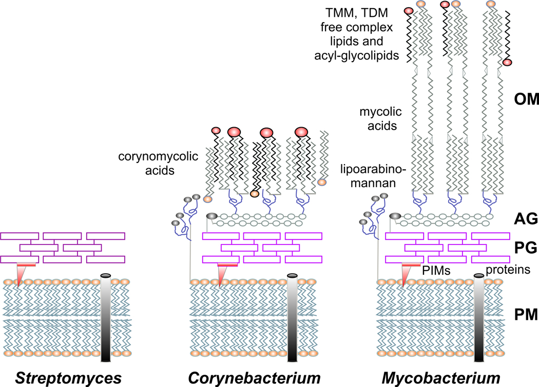
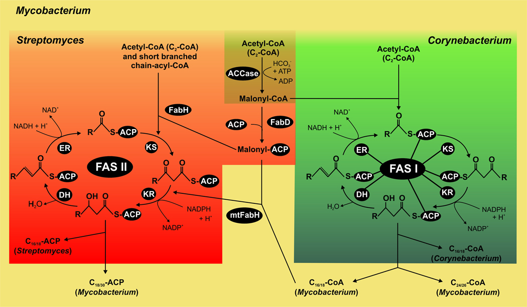
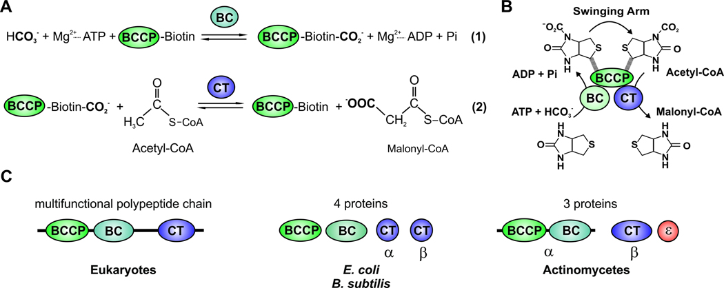
All organisms that produce fatty acids do so via a repeated cycle of reactions. In mammals and other animals, these reactions are catalyzed by a type I fatty acid synthase (FAS), a large multifunctional protein to which the growing chain is covalently attached. In contrast, most bacteria (and plants) contain a type II system in which each reaction is catalyzed by a discrete protein. The pathway of fatty acid biosynthesis in Escherichia coli is well established and has provided a foundation for elucidating the type II FAS pathways in other bacteria (White et al., 2005). However, fatty acid biosynthesis is more diverse in the phylum Actinobacteria: Mycobacterium, possess both FAS systems while Streptomyces species have only the multienzyme FAS II system and Corynebacterium species exclusively FAS I. In this review, we present an overview of the genome organization, biochemical properties and physiological relevance of the two FAS systems in the three genera of actinomycetes mentioned above. We also address in detail the biochemical and structural properties of the acyl-CoA carboxylases (ACCases) that catalyzes the first committed step of fatty acid synthesis in actinomycetes, and discuss the molecular bases of their substrate specificity and the structure-based identification of new ACCase inhibitors with antimycobacterial properties.


Figures

Fig. 1
Fig. 1. Schematic representation of the cell envelope of actinomycetes
Comparison of the cell wall structures from Streptomyces, Corynebacterium and Mycobacterium. In streptomycetes, as in other Gram-positive bacteria, the cell wall consists of a peptidoglycan layer (PG) that covers the cytoplasmic membrane (PM). Corynebacteria and mycobacteria contain a more complex cell wall including an additional layer of arabinogalactan (AG) and an outer membrane (OM) composed of mycolic acids, trehalose monomycolate (TMM), trehalose dimycolate (TDM) and free lipids. The mycobacterial cell wall has a high proportion of covalently attached mycolic acid residues, which constitute a permeability barrier contributing to antibiotic resistance and pathogenicity. PIMs: phosphatidylinositol-mannosides.
Fig. 2
Fig. 2. Comparative model of fatty acid biosynthesis in actinomycetes
Streptomyces Corynebacterium and Mycobacterium share the initiation step for fatty acid biosynthesis which is the carboxylation of acetyl-CoA to produce malonyl-CoA, catalyzed by the ACCase complex (brown). Both in Mycobacterium and Corynebacterium spp., malonyl-CoA and acetyl-CoA are condensed to be utilized by the multifunctional type I FAS for de novo biosynthesis of fatty acids (green). In Streptomyces, malonyl-CoA is converted to malonyl-ACP by FabD and then condensed with acetyl-CoA by FabH to form β-ketoacyl-ACP, which is used by the type II FAS dissociated system for de novo biosynthesis of fatty acids (red). Both in FAS I and FAS II systems, the chain elongation steps consist of an iterative series of reactions built on successive addition of a two-carbon unit to a nascent acyl group, and reaction intermediates are covalently attached to the acyl carrier protein (ACP). In Mycobacterium both systems are present, FAS I for de novo fatty acid biosynthesis and FAS II for the elongation of fatty acids for mycolic acid production. The differential features of Mycobacterium are shown in yellow.
Fig. 3
Fig. 3. Genetic organization of FAS II genes
Synteny present in the fas II (fab) operons (A), the cluster of genes encoding for the FAS-II reductases (B) and the FAS II dehydratases (C) in some of the most relevant species of mycobacteria and streptomycetes: M. tuberculosis (Mt), M. smegmatis (Ms), S. coelicolor (Sc), S. griseous (Sg) and S. avermitilis (Sa).
Fig. 4
Fig. 4. Schematic diagram of ACC reaction and of domains and/or subunits of the biotin-dependent ACC
(A) Stepwise enzymatic reactions of ACC. (B) ACC is composed of three different components, the biotin carboxylase component (BC), the biotin carboxyl carrier protein (BCCP) and carboxyltransferase (CT). The “Swinging Arm” of biotin-BCCP transports CO2 between the components BC and CT. Then the carboxyl group is transferred from biotin to acetyl-CoA to form malonyl-CoA. (C) The eukaryotic ACC is a multifunctional polypeptide chain that contains the three components (BC, BCCP and CT). Bacterial ACC is composed of multiple subunits. The E. coli and B. subtilis ACCs are formed by four proteins: the subunit BC, the subunit BCCP, and two proteins (α and β) that form the CT subunit. In actinomycetes the ACCase complex is made up of two main subunits: the α subunit, which contains the BC and BCCP components and the β subunit, which has the CT activity. In addition, actinomycete ACCases may include a third subunit, ε, the presence of which dramatically stimulates the specific activity of the enzyme complexes.
Fig. 5
Fig. 5. Phylogenetic analysis and genetic organization of CTs β subunits of ACCase complexes found in actinomycetes
The unrooted phylogenetic tree was generated using the neighbor-joining method of the freely available Molecular Evolutionary Genetics Analysis (MEGA) software version 4.0. Reliability of the tree was assessed by bootstrap analysis with 1,000 replications. Bootstrap values for the different families described in the text are all over 80. The tree is drawn to scale and the lengths of the branches are proportional to the inferred evolutionary distances. The bar indicates the relative number of substitutions per site. The organism key is as follows: Ce, Corynebacterium efficiens, Cg, Corynebacterium glutamicum; DIP, Corynebacterium diphtheriae; Mb, Mycobacterium bovis; Ml, Mycobacterium leprae; Mt, Mycobacterium tuberculosis; Ms, Mycobacterium smegmatis; PPA, Propionibacterium acnes; Sa, Streptomyces antibioticus; SAV, Streptomyces avermitilis; SCO, Streptomyces coelicolor; SGR, Streptomyces griseus; Sv, Streptomyces venezuelae; Tfu, Thermobifida fusca. The genetic organization and synteny are shown for CTs that have been characterized (Mt AccD4, Mt AccD5, Cg accD1, Cg AccD2, Cg AccD3, SCO PccB, and SCO AccB).
Fig. 6
Fig. 6. Crystal structure of PccB from S. coelicolor
(A) The overall hexameric structure of PccB formed by two stacks of three monomers (A-B-C and D-E-F) related by the 3-fold axis. (B) Monomer A of PccB, colored from the N-terminus (blue) to the C-terminus (red), with two structurally similar domains (N and C). Monomer D is shown in black and white under monomer A. (C) The dimeric, di-domain interaction between monomer A and monomer D (after a 90° rotation) of PccB, related by the 2-fold axis. (D) Electronegative surfaces of AccB, PccB, and 12S (Diacovich et al., 2004; Hall et al., 2003) reveal key differences between these very similar CTs. Surfaces are colored according to their charges from red (−) to white to blue (+).
Fig. 7
Fig. 7. PccB active site
(A) The binding pockets of acyl-CoA and biotin in PccB are perpendicular to each other and meet at the junction of the L-shaped active site. (B) The binding pocket of propionyl-CoA reveals that residue 422, at the end of the pocket, is the only residue different in AccB and PccB. (C) Sequence comparison of different CTs that accept propionyl-CoA as a substrate, like PccB, where position 422 (in PccB) is occupied by small residues (blue star), such as Asp or Cys, and CTs that accept acetyl-CoA as a substrate, such as AccB, where this residue is a larger, hydrophobic residue. Scoel (S. coelicolor), Seryt (S. erythraea), Mtub (M. tuberculosis), Hum (Human), Yeast, Svene (S. venezuelae), Tteng (T. tengcongensis).
Fig. 8
Fig. 8. Crystal structure of AccD5 from M. tuberculosis
(A) The overall hexameric fold of AccD5 is similar to that of PccB. (B) The active site lies at the dimeric, di-domain interface. (C) A comparison of the active sites between PccB and AccD5 shows that AccD5 cannot accommodate a substrate larger than propionyl-CoA (Lin et al., 2006)

References

    1. Alberts AW, Nervi AM, Vagelos PR. Acetyl CoA carboxylase, II. Demonstration of biotin-protein and biotin carboxylase subunits. Proc Natl Acad Sci U S A. 1969;63:1319–1326. - PMC - PubMed
    1. Alberts AW, Bell RM, Vagelos PR. Acyl carrier protein. XV. Studies of - ketoacyl-acyl carrier protein synthetase. J Biol Chem. 1972;247:3190–3198. - PubMed
    1. Anderson AC. The process of structure-based drug design. Chem Biol. 2003;10:787–797. - PubMed
    1. Arabolaza A, Rodriguez E, Altabe S, Alvarez H, Gramajo H. Multiple pathways for triacylglycerol biosynthesis in Streptomyces coelicolor. Appl Environ Microbiol. 2008;74:2573–2582. - PMC - PubMed
    1. Arabolaza A, D'Angelo M, Comba S, Gramajo H. FasR, a novel class of transcriptional regulator, governs the activation of fatty acid biosynthesis genes in Streptomyces coelicolor. Mol Microbiol. 2010a;78:47–63. - PubMed

Publication types

MeSH terms