. 2010 Dec 24;330(6012):1816-20.
doi: 10.1126/science.1195821.

Structures of C3b in complex with factors B and D give insight into complement convertase formation

Affiliations

Structures of C3b in complex with factors B and D give insight into complement convertase formation

Federico Forneris et al. Science. .

Abstract

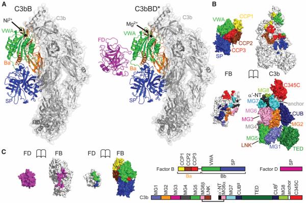
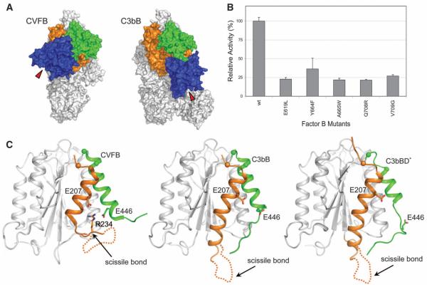
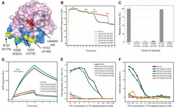
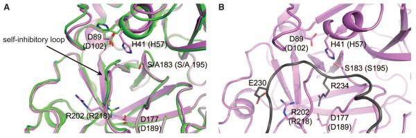
Activation of the complement cascade induces inflammatory responses and marks cells for immune clearance. In the central complement-amplification step, a complex consisting of surface-bound C3b and factor B is cleaved by factor D to generate active convertases on targeted surfaces. We present crystal structures of the pro-convertase C3bB at 4 angstrom resolution and its complex with factor D at 3.5 angstrom resolution. Our data show how factor B binding to C3b forms an open "activation" state of C3bB. Factor D specifically binds the open conformation of factor B through a site distant from the catalytic center and is activated by the substrate, which displaces factor D's self-inhibitory loop. This concerted proteolytic mechanism, which is cofactor-dependent and substrate-induced, restricts complement amplification to C3b-tagged target cells.


Figures

Fig. 1
Fig. 1
Structures of C3bB and C3bBD*. (A) Overall structure of C3bB (left) and C3bBD* (right). C3b is shown as gray transparent surface; FB is shown as orange (Ba), green (VWA), and blue (SP) cartoons; and FD is represented as a magenta cartoon. The black spheres highlight the metal ions (Ni2+ for C3bB, Mg2+ for C3bBD*) at the MIDAS site. (B) Opened view of the footprint of the C3b-FB interaction, highlighting the domains of FB on the C3b surface (top) and the domains of C3b on FB (bottom). (C) Opened view of the footprint of the FB-FD interaction, highlighting the domains of FB on the FD surface (left) and the single domain of FD on FB (right). The scheme indicates the domain compositions and color codes of C3b, FB, and FD used in (B) and (C).
Fig. 2
Fig. 2
Comparison between the closed and open states in the pro-convertase C3bB. (A) Surface representation of the closed (CVFB, left) (16) and open (C3bB, right) conformations of the pro-convertase. Colors are the same as in Fig. 1A. Red triangles indicate the position of the catalytic site of FB during the conformational changes. (B) FD-mediated cleavage of C3bB performed by using SDS–polyacrylamide gel electrophoresis (PAGE) shows that mutations (28) in FB located at the interface between the SP and CUB domains in the open C3bB pro-convertase lower convertase formation rates. The histogram shows the relative pro-convertase activation rates compared with those of wild-type FB (see also fig. S6). Error bars represent deviations from the mean observed in multiple experiments (n>3). (C) Cartoon diagram of the VWA domain of FB, highlighting conformational changes in the transition from the closed (left) to the open state of the pro-convertase in absence (center) or in presence of FD (right). The αL helix is colored in orange and the α7 helix in green. The putative orientation of the loop containing the scissile bond of FB is shown with a dashed line. The positions of the C-α atoms located at the N-termini of αL and α7 helix are shown as spheres.
Fig. 3
Fig. 3
Analysis of FD exosite. (A) Surface diagram of FD highlighting its exosite in blue and its catalytic site in red (28). Exosite mutations are shown in yellow, whereas the biotinylation site associated with FD inactivation (26) is shown in green. (B) FD-mediated cleavage of C3bB pro-convertases monitored by SPR; FD mutants were injected onto surface-bound C3bB, and the cleavage activity was compared with that of wild type (wt). A drop in SPR response upon FD injection corresponds to the removal of the Ba fragment. Wild-type FD was injected at the end of each experiment to ensure cleavage sensitivity. (C) FD-mediated cleavage of C3bB performed by using SDS-PAGE with FD exosite mutants. The histogram shows the relative pro-convertase activation rates compared with those of wild-type FD. Error bars represent deviations from the mean observed in multiple experiments (n>3). (D) Effect of FD exosite mutations on the formation and decay of the C3bBb convertase monitored by SPR; wild-type FB was premixed with various FD mutants and injected over immobilized C3b. RU, resonance units. (E) Reconstitution of complement activity in FD-depleted plasma using FD mutants determined as C3b deposition on the enzyme-linked immunosorbent assay (ELISA) plate. O.D., optical density. (F) Hemolytic activity assays using FD-depleted plasma reconstituted with different mutants of FD: Lysis of rabbit erythrocytes was monitored by colorimetry and compared with 100% haemolysis in water.
Fig. 4
Fig. 4
Analysis of FD catalytic site. (A) Conformational changes observed in FD catalytic site in C3bBD* structure. Superposition of the structure of FD S183A (S195A) from C3bBD* (magenta) with wild-type free FD (green, PDB ID 1DSU) (22, 28) showing the displacement of the self-inhibitory loop and flipping of the side chain of His41 (His57) to the catalytic conformation. (B) Zoomed view of FD catalytic site with modeled FB scissile bond loop bound (dark gray). The model highlights the putative interaction between Glu230 of FB and Arg202 (Arg218) of FD and the P1 residue Arg234 making a salt bridge with Asp177 (Asp189).

References

    1. Ricklin D, Hajishengallis G, Yang K, Lambris JD. Nat. Immunol. 2010;11:785. - PMC - PubMed
    1. Walport MJ. N. Engl. J. Med. 2001;344:1058. - PubMed
    1. Pangburn MK, Müller-Eberhard HJ. Biochem. J. 1986;235:723. - PMC - PubMed
    1. Gros P, Milder FJ, Janssen BJ. Nat. Rev. Immunol. 2008;8:48. - PubMed
    1. Xu Y, Narayana SV, Volanakis JE. Immunol. Rev. 2001;180:123. - PubMed

Publication types

MeSH terms

LinkOut - more resources