

Thalamofrontal neurodevelopment in new-onset pediatric idiopathic generalized epilepsy
- PMID: 21205692
- PMCID: PMC3030227
- DOI: 10.1212/WNL.0b013e318203e8f3
Thalamofrontal neurodevelopment in new-onset pediatric idiopathic generalized epilepsy
Abstract
Background: Quantitative MRI techniques have demonstrated thalamocortical abnormalities in idiopathic generalized epilepsy (IGE). However, there are few studies examining IGE early in its course and the neurodevelopmental course of this region is not adequately defined.
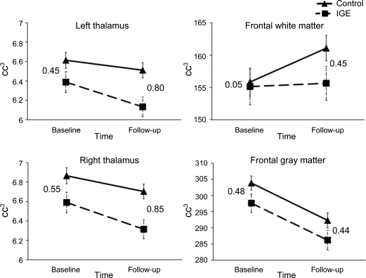
Objective: We examined the 2-year developmental course of the thalamus and frontal lobes in pediatric new-onset IGE (i.e., within 12 months of diagnosis).
Methods: We performed whole-brain MRI in 22 patients with new-onset IGE and 36 age-matched healthy controls. MRI was repeated 24 months after baseline MRI. Quantitative volumetrics were used to examine thalamic and frontal lobe volumes.
Results: The IGE group showed significant differences in thalamic volume within 1 year of seizure onset (baseline) and went on to show thalamic volume loss at a significantly faster rate than healthy control children over the 2-year interval. The control group also showed a significantly greater increase in frontal white matter expansion than the IGE group. In contrast, frontal lobe gray matter volume differences were moderate at baseline and persisted over time, indicating similar developmental trajectories with differences early in the disease process that are maintained.
Conclusions: Brain tissue abnormalities in thalamic and frontal regions can be identified very early in the course of IGE and an abnormal trajectory of growth continues over a 2-year interval.
Figures
References
-
- Mattson RH. Overview: idiopathic generalized epilepsies. Epilepsia 2003;44:2–6 - PubMed
-
- Hommet C, Sauerwein HC, De Toffol B, Lassonde M. Idiopathic epileptic syndromes and cognition. Neurosci Biobehav Rev 2006;30:85–96 - PubMed
-
- Gloor P, Pellegrini A, Kostopoulos GK. Effects of changes in cortical excitability upon the epileptic bursts in generalized penicillin epilepsy of the cat. Electroencephalogr Clin Neurophysiol 1979;46:274–289 - PubMed
-
- Bernhardt BC, Rozen DA, Worsley KJ, Evans AC, Bernasconi N, Bernasconi A. Thalamo-cortical network pathology in idiopathic generalized epilepsy: insights from MRI-based morphometric correlation analysis. Neuroimage 2009;46:373–381 - PubMed
-
- Deppe M, Kellinghaus C, Duning T, et al. Nerve fiber impairment of anterior thalamocortical circuitry in juvenile myoclonic epilepsy. Neurology 2008;71:1981–1985 - PubMed