. 2011 Feb;138(3):507-18.
doi: 10.1242/dev.060939.

C. elegans bicd-1, homolog of the Drosophila dynein accessory factor Bicaudal D, regulates the branching of PVD sensory neuron dendrites

Affiliations

C. elegans bicd-1, homolog of the Drosophila dynein accessory factor Bicaudal D, regulates the branching of PVD sensory neuron dendrites

Cristina Aguirre-Chen et al. Development. 2011 Feb.

Abstract

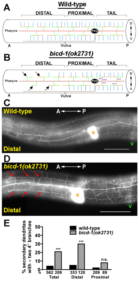
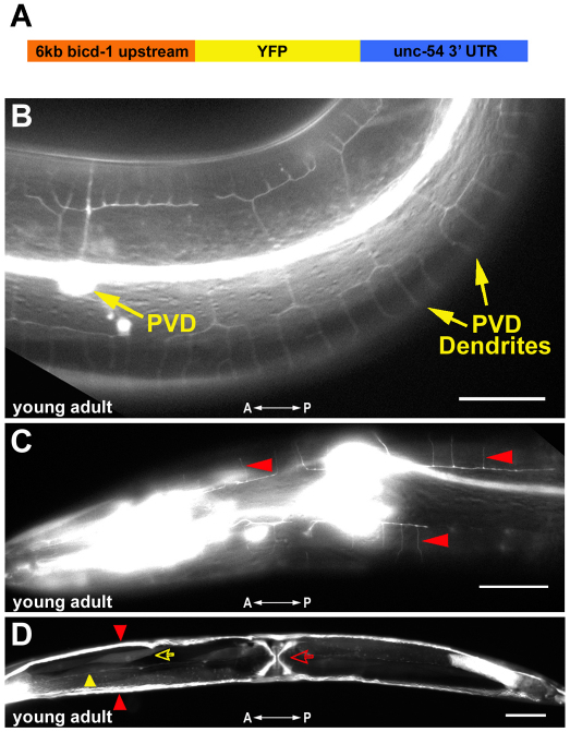
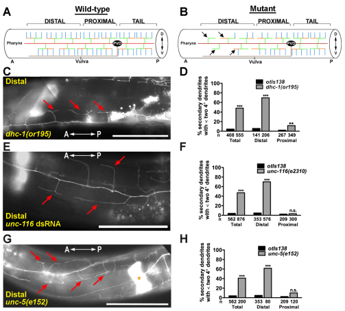
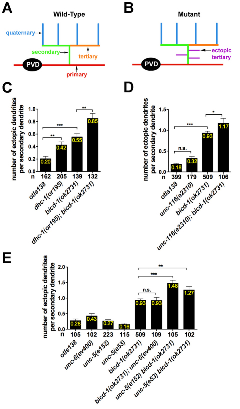
The establishment of cell type-specific dendritic arborization patterns is a key phase in the assembly of neuronal circuitry that facilitates the integration and processing of synaptic and sensory input. Although studies in Drosophila and vertebrate systems have identified a variety of factors that regulate dendrite branch formation, the molecular mechanisms that control this process remain poorly defined. Here, we introduce the use of the Caenorhabditis elegans PVD neurons, a pair of putative nociceptors that elaborate complex dendritic arbors, as a tractable model for conducting high-throughput RNAi screens aimed at identifying key regulators of dendritic branch formation. By carrying out two separate RNAi screens, a small-scale candidate-based screen and a large-scale screen of the ~3000 genes on chromosome IV, we retrieved 11 genes that either promote or suppress the formation of PVD-associated dendrites. We present a detailed functional characterization of one of the genes, bicd-1, which encodes a microtubule-associated protein previously shown to modulate the transport of mRNAs and organelles in a variety of organisms. Specifically, we describe a novel role for bicd-1 in regulating dendrite branch formation and show that bicd-1 is likely to be expressed, and primarily required, in PVD neurons to control dendritic branching. We also present evidence that bicd-1 operates in a conserved pathway with dhc-1 and unc-116, components of the dynein minus-end-directed and kinesin-1 plus-end-directed microtubule-based motor complexes, respectively, and interacts genetically with the repulsive guidance receptor unc-5.


Figures

Fig. 1.
Fig. 1.
Spatiotemporal pattern of PVD dendrite branching. (A-F) The otIs138 transcriptional reporter was used to characterize the branching of PVD dendrites in the tail region, posterior to the PVD cell body, of wild-type worms at the L2 (A-C; L2′, L2′ and L2′′ refer to progressively later stages of development in L2 animals), L3 (D) and L4 (E) larval stages and at the young adult stage (F). Schematic representations of reporter labeling of PVD dendritic arbors are shown together with low (A,D-F) and high (A-F) magnification images of examples of the schematized projections. Red boxes in A and D-F represent the area shown in the high magnification image. Anteroposterior orientation is indicated by the white double-headed arrow. Asterisk in F denotes the spermatheca. The labeling of only one (left or right) PVD neuron and one of its dendritic trees is depicted in each schematic, and labeled PVD neuron cell bodies are identified by arrows in each of the middle panels. Scale bars: 10 μm.
Fig. 2.
Fig. 2.
RNAi constructs fed to wild-type worms efficiently knock down gene expression in PVD neurons. (A,B,D) otIs138 animals fed the empty vector construct, L4440, display minimal knockdown of GFP protein expression in PVD neurons (A); 96% of animals retain GFP expression in both PVD cell bodies (D). The green and red arrows in A point to labeled PVD and PDE cell bodies, respectively. otIs138 animals fed GFP dsRNA exhibit reduced GFP protein expression in PVD neurons (B); 28.3% of animals exhibit GFP expression in both PVD cell bodies, 41.3% express GFP in one PVD cell body only and 30.4% exhibit no GFP expression in both PVD cell bodies (D). The green arrow in B indicates the absence of GFP protein expression in a location presumably occupied by the PVD neuron. Because diminished GFP expression within PVD neurons prevented the visualization of PVD dendritic arbors (data not shown) in an RNAi hypersensitive strain (Schmitz et al., 2007), we conducted the complete screen in an otherwise wild-type background. (C,E) Half of otIs138 animals fed mec-3 dsRNA (C) display a significant reduction in the numbers of secondary, tertiary and quaternary dendrites (E), phenocopying the branch-reducing phenotype exhibited by mec-3(u298) genetic mutants. Anteroposterior orientation is indicated by the white double-headed arrow. The asterisks in A and C mark the spermatheca. Scale bars: 50 μm. Error bars represent s.e.m. ***P<0.001 determined by Fisher's exact test.
Fig. 3.
Fig. 3.
bicd-1(ok2731) animals phenocopy the bicd-1 RNAi enhanced-branching defect. (A) Genetic position of bicd-1 on chromosome IV and its exon-intron structure, with the positions of the ok2731 and tm3421 mutations indicated. (B,C) Schematics of individual PVD dendrite trees present in wild-type (B) and bicd-1(ok2731) or bicd-1 RNAi-treated (C) animals. (D,E) otIs138 animals fed bicd-1 dsRNA exhibit an enhanced-branching phenotype posterior to the PVD cell body (D). bicd-1(ok2731) animals phenocopy the bicd-1 RNAi enhanced-branching phenotype (E). Purple arrowheads indicate ectopic tertiary dendrites. Asterisks indicate the spermatheca. Anteroposterior orientation is indicated by the white double-headed arrow. (F) Quantification of the enhanced-branching phenotype exhibited by otIs138 young adult animals subjected to bicd-1 RNAi. (G) The PVD enhanced-branching phenotype is first apparent at the young adult stage. n, number of secondary dendrites scored in F and G. Error bars represent s.e.m. ***P<0.001 determined by unpaired Student's t-test with Bonferroni adjustment. n.s., not significant. Scale bars: 10 μm.
Fig. 4.
Fig. 4.
bicd-1(ok2731) animals exhibit a reduction in the number of dendritic branches distal to the PVD cell body. (A,B) Schematics of the wild-type (A) and bicd-1(ok2731) (B) PVD arborization pattern. `Distal' specifies the region of the PVD arbor anterior to the vulva and `proximal' denotes the region between the vulva and the PVD cell body. Arrows indicate individual dendritic trees that possess less than two quaternary dendrites. Dorsoventral orientation is indicated by the double-headed arrow. (C,D) Images of the distal regions of wild-type (C) and bicd-1(ok2731) (D) PVD arbors of young adult animals (52-58 hours post hatch, room temperature). Asterisks mark the spermatheca and `v' indicates the position of the vulva. Red arrows point to individual dendritic trees that possess less than two quaternary dendrites. (E) Quantification of the branch-reducing phenotype in both the distal and proximal regions of the PVD arbor. n, number of secondary dendrites scored. Error bars represent s.e.m. ***P<0.001 determined by chi-square test with Bonferroni adjustment. n.s., not significant. Scale bars: 50 μm.
Fig. 5.
Fig. 5.
bicd-1::YFP expression pattern. (A) A bicd-1 transcriptional reporter construct was generated by fusing 6kb of genomic sequence directly upstream of the bicd-1 ATG to the yfp/unc-54 3′UTR cassette. (B) Yellow arrows mark YFP protein expression detected in the PVD neuronal cell body and its dendrites at the young adult stage. (C) Red arrowheads point to labeled quaternary dendrites of FLP neurons in the head. (D) Ventral view of a bicd-1::YFP labeled animal. The red and yellow open arrows point to labeled vulva and body wall muscles, respectively. Red arrowheads identify YFP protein expression in the bilaterally symmetrical excretory canals and the yellow arrowhead points to YFP protein expression in the posteriorly directed processes of AVF neurons. Anteroposterior orientation is indicated by the white double-headed arrow. Scale bars: 25 μm in B and C; 50 μm in D.
Fig. 6.
Fig. 6.
bicd-1 acts in PVD neurons. (A) bicd-1 cDNA, expressed under the control of either the PVD specific promoter ser2prom3 or the muscle-specific promoter myo-3, partially rescues the enhanced-branched phenotype. (B) Mosaic analysis was conducted using a C43G2 cosmid rescuing line that exhibited complete rescue of the enhanced-branching phenotype (rzEx111, see Fig. S1 in the supplementary material). mec-7::gfp and myo-3::rfp were used to mark the AB.p and P1 lineages, respectively. b.w. muscle, body wall muscle. (C) Quantification of the enhanced-branching phenotype present in mosaic animals demonstrates that bicd-1 acts in PVD neurons. [AB.p+P1] denotes animals that have retained the array in both the AB and P1 lineages. [AB.p-] and [P1-] denote animals that have retained the array in either the P1 or AB.p lineage, respectively. Adult animals scored in the mosaic analyses were initially selected at the L4 stage and scored for rescue 24 hours later. n, number of secondary dendrites scored. Error bars represent s.e.m. **P<0.01 determined by one-way ANOVA with Dunnett's post test.
Fig. 7.
Fig. 7.
dhc-1(or195), unc-116(e2310) and unc-5(e152) mutants possess fewer dendrite branches distal to the PVD cell body. (A,B) Schematics of the wild-type (A) and mutant (B) PVD arborization pattern. `Distal' specifies the region of the PVD arbor anterior to the vulva and `proximal' denotes the region between the vulva and the PVD cell body. Arrows point to individual dendritic trees that possess less than two quaternary dendrites. (C,E,G) Micrographs of the distal regions of dhc-1(or195) (C), unc-116 RNAi (E) and unc-5(e152) (G) PVD arbors. Red arrows point to the absence of quaternary dendrites. The asterisk in G indicates the spermatheca. Anteroposterior orientation is indicated by the white double-headed arrow. (D,F,H) Quantification of the branch-reducing phenotype in the distal and proximal regions of the PVD arbor at 25°C (D) and 20°C (F,H). n, number of secondary dendrites scored. dhc-1(or195) mutants are temperature sensitive and all analyses utilizing this allele were performed at 25°C. Scale bars: 50 μm. Error bars represent s.e.m. **P<0.01, ***P<0.001 determined by chi-square test with Bonferroni adjustment. n.s., not significant.
Fig. 8.
Fig. 8.
bicd-1 genetically interacts with dhc-1, unc-116 and unc-5 to regulate PVD dendrite branching. (A,B) Schematic depictions of individual PVD arbors present in wild-type (A) and mutant (B) animals. (C-E) Quantification of the enhanced-branching phenotype posterior to the PVD cell body (tail region) at 25°C (C) and 20°C (D,E). n, number of secondary dendrites scored. Error bars represent s.e.m. *P<0.05, **P<0.01, ***P<0.001 determined by one-way ANOVA with Dunnett's post test. n.s., not significant.

References

    1. Ashrafi K., Chang F. Y., Watts J. L., Fraser A. G., Kamath R. S., Ahringer J., Ruvkun G. (2003). Genome-wide RNAi analysis of Caenorhabditis elegans fat regulatory genes. Nature 421, 268-272 - PubMed
    1. Baens M., Marynen P. (1997). A human homologue (BICD1) of the Drosophila bicaudal-D gene. Genomics 45, 601-606 - PubMed
    1. Brenner S. (1974). The genetics of Caenorhabditis elegans. Genetics 77, 71-94 - PMC - PubMed
    1. Bullock S. L., Ish-Horowicz D. (2001). Conserved signals and machinery for RNA transport in Drosophila oogenesis and embryogenesis. Nature 414, 611-616 - PubMed
    1. Bullock S. L., Nicol A., Gross S. P., Zicha D. (2006). Guidance of bidirectional motor complexes by mRNA cargoes through control of dynein number and activity. Curr. Biol. 16, 1447-1452 - PubMed

Publication types

MeSH terms

LinkOut - more resources