. 2011 Jan 25;108(4):1421-6.
doi: 10.1073/pnas.1011784108. Epub 2011 Jan 4.

Direct evidence for functional smooth muscle myosin II in the 10S self-inhibited monomeric conformation in airway smooth muscle cells

Affiliations

Direct evidence for functional smooth muscle myosin II in the 10S self-inhibited monomeric conformation in airway smooth muscle cells

Deanna L Milton et al. Proc Natl Acad Sci U S A. .

Abstract

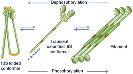
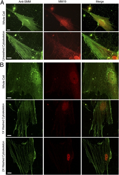
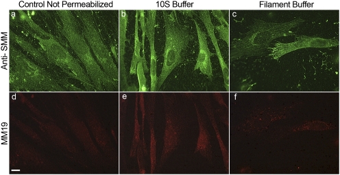
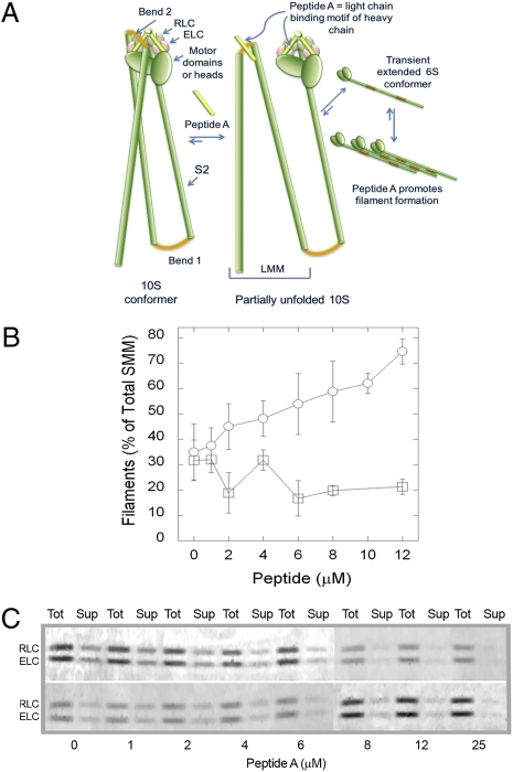
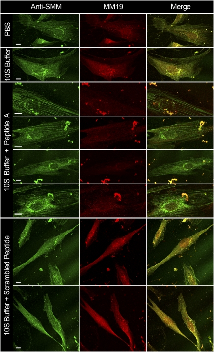
The 10S self-inhibited monomeric conformation of myosin II has been characterized extensively in vitro. Based upon its structural and functional characteristics, it has been proposed to be an assembly-competent myosin pool in equilibrium with filaments in cells. It is known that myosin filaments can assemble and disassemble in nonmuscle cells, and in some smooth muscle cells, but whether or not the disassembled pool contains functional 10S myosin has not been determined. Here we address this question using human airway smooth muscle cells (hASMCs). Using two antibodies against different epitopes on smooth muscle myosin II (SMM), two distinct pools of SMM, diffuse, and stress-fiber-associated, were visualized by immunocytochemical staining. The two SMM pools were functional in that they could be interconverted in two ways: (i) by exposure to 10S- versus filament-promoting buffer conditions, and (ii) by exposure to a peptide that shifts the filament-10S equilibrium toward filaments in vitro by a known mechanism that requires the presence of the 10S conformation. The effect of the peptide was not due to a trivial increase in SMM phosphorylation, and its specificity was demonstrated by use of a scrambled peptide, which had no effect. Based upon these data, we conclude that hASMCs contain a significant pool of functional SMM in the 10S conformation that can assemble into filaments upon changing cellular conditions. This study provides unique direct evidence for the presence of a significant pool of functional myosin in the 10S conformation in cells.


Conflict of interest statement

The authors declare no conflict of interest.

Figures

Fig. 1.
Fig. 1.
Schematic of conformational states of SMM derived from in vitro studies. Green, tail with red bending regions and globular heads; brown and pink represent ELC, and RLC, respectively (Fig. 4 gives a more detailed description of 10S structure). Equilibration between filaments and 10S likely proceeds through the transient extended monomeric 6S conformer. Phosphorylation at S19 on RLC promotes filament assembly and dephosphorylation promotes disassembly of filaments to 10S. Only one head is shown on the filament for clarity. In both the filament and 6S schematics, the heads are drawn to the same scale as in the 10S schematic, but the tails are drawn to a smaller scale, making them about three times shorter than they actually are.
Fig. 2.
Fig. 2.
Visualization of SMM pools in contractile phenotype cells. (A) Untreated whole (63×) versus Triton-permeabilized cells (cytoskeletons; 100×) that were not washed. Anti-total SMM stain (green), MM19 stain (red), and merged images (Right). Controls without primary antibodies were black. (B) Confocal images (100×) showing the effect of washing the cytoskeletons with PBS without ATP. (Top Row) Whole cell, middle cytoskeleton prepared as in Materials and Methods but further washed with PBS one time (Middle Row) and two times (Bottom Row). (All scale bars, 10 μm.)
Fig. 3.
Fig. 3.
Effect of buffer conditions on SMM pools in α-toxin–permeabilized confluent proliferating cells. Green represents anti-SMM; red, MM19 antibody. Control not permeabilized (A, D); α-toxin permeabilized and exposed to 10S buffer (B, E) and α-toxin permeabilized exposed to filament buffer (C, F). (Scale bar, 10 μm.) Images D, E, and F were despeckled using National Institutes Health ImageJ software to reduce camera noise.
Fig. 4.
Fig. 4.
Peptide A mechanism and effects on filament-monomer equilibrium in vitro. (A) Schematic showing mechanism of inhibition of SMM hairpin bend (Bend 2) interaction with the light chain region by Peptide A. Green indicates HC including the globular motor domains, the S2 descending region of the tail, and the light meromyosin (LMM) region of the tail; red, bends in HC; pink, ELC; and brown, RLC. The 10S conformer (left) is fully bent with Bend 2 in LMM interacting with heads. Cartoon is partly based upon structural data from Seow (8) and Salzameda et al. (24). Peptide A, which is identical in sequence to the light chain binding motif of the HC (light green), will compete with that region for interaction with Bend 2, stabilizing the 6S conformer and promoting filaments. (B) Effects of synthetic peptides on equilibrium between filaments and soluble SMM in vitro. Chicken gizzard SMM (1 mg/mL) with Peptide A (○) or scrambled peptide (□) in 1 mM MgCl2, 150 mM NaCl, 10 mM Imidazole (pH 7.0), 0.1 mM EGTA, 0.1 mM DTT, and 1 mM ATP. The filaments were pelleted by ultracentrifugation, and the amount of soluble and total SMM was determined by a Bradford assay. Data represent the results from three independent experiments on two different SMM preparations. Error bars represent SEM. (C) Images of the light chain region from Coomassie-stained SDS gels of samples from two different (Upper and Lower) in vitro Peptide A titrations using equal loading volumes of the sample before (Tot) and after (Sup) pelleting filaments using the same protocol as in B.
Fig. 5.
Fig. 5.
Effect of synthetic peptides on nonconfluent proliferating hASMCs. First column, hASMCs stained with anti-SMM; second column, stained with MM19; third column, merged. Row labels indicate treatments. PBS, control not permeabilized, exposed to PBS; 10S buffer, α-toxin–permeabilized exposed to 10S buffer; 10S Buffer + Peptide A or scrambled peptide, exposed to 30 μM Peptide A or scrambled peptide, in 10S buffer. Bright particles are cell debris. All images were taken on a confocal microscope, 63× or 100×. (All scale bars, 10 μm.)

References

    1. Cremo CR, Hartshorne DJ. In: Myosins: A Superfamily of Molecular Motors. Coluccio LM, editor. Vol. 7. Dordrecht, Netherlands: Springer; 2008. pp. 171–222.
    1. Onishi H, Wakabayashi T. Electron microscopic studies of myosin molecules from chicken gizzard muscle I: The formation of the intramolecular loop in the myosin tail. J Biochem. 1982;92:871–879. - PubMed
    1. Trybus KM, Huiatt TW, Lowey S. A bent monomeric conformation of myosin from smooth muscle. Proc Natl Acad Sci USA. 1982;79:6151–6155. - PMC - PubMed
    1. Kendrick-Jones J, Smith RC, Craig R, Citi S. Polymerization of vertebrate non-muscle and smooth muscle myosins. J Mol Biol. 1987;198:241–252. - PubMed
    1. Trybus KM, Lowey S. Conformational states of smooth muscle myosin. Effects of light chain phosphorylation and ionic strength. J Biol Chem. 1984;259:8564–8571. - PubMed

Publication types

MeSH terms

LinkOut - more resources