. 2011 Jan 25;108(4):1549-54.
doi: 10.1073/pnas.1017340108. Epub 2011 Jan 4.

T-cell receptor ligation induces distinct signaling pathways in naive vs. antigen-experienced T cells

Affiliations

T-cell receptor ligation induces distinct signaling pathways in naive vs. antigen-experienced T cells

Keishi Adachi et al. Proc Natl Acad Sci U S A. .

Abstract

Naïve T lymphocytes display weaker and slower responses than antigen-experienced cells for reasons that are not well understood. Here we show that T-cell receptor (TCR) stimulation induces distinct ERK and p38 phosphorylation patterns in naïve and antigen-experienced human T cells, and that these contribute to the differential responses shown by these cells. Specifically, TCR ligation triggers the activation of the ERK pathway in naïve cells. This phosphorylation of ERK attenuates subsequent calcium influx and accelerates the degradation of the signalsome. In contrast, anti-CD3 stimulation of experienced cells results in the phosphorylation of p38 via an association with Discs large (Dlg). Thus, there are distinct signaling pathways triggered by TCR ligation that impair signaling in naïve cells and facilitate it in antigen-experienced cells.


Conflict of interest statement

The authors declare no conflict of interest.

Figures

Fig. 1.
Fig. 1.
ERK phosphorylation is induced preferentially in CD4+ naïve cells upon TCR stimulation. (A and B) ERK phophorylation (ppERK) in human PBMCs was analyzed by Phosphoflow. Stimulations were done with 10 μg/mL anti-CD3ε (S4.1) and/or 5 μg/mL anti-CD28 (αCD3 + αCD28 crosslink). (A) Cells were stimulated for 0, 2, or 5 min, and CD4+ T-cell population (the unfractionated CD4+ population) was gated using Flowjo. (B) The CD4+ T-cell population was stimulated for 2 min and divided into antigen-naïve (red) and -experinced (blue) populations by CD45RA or CD45RO expression, respectively. (C and D) ERK phosphorylation levels of naïve and antigen-experienced populations were compared with normalized values. Values were calculated as follows: (phosphoprotein-expressing cell percentage of each population)/(that of unfractionated CD4+ population). Stimulation time was 2 min, and anti-CD3ε (S4.1) (C) or anti-CD3γ (UCHT1) (D) were used. Data represent the mean + SD of seven (C) or three (D) independent experiments.
Fig. 2.
Fig. 2.
p38 is phosphorylated predominantly in experienced CD4+ T cells as a result of TCR stimulation. (A and B) Phosphoflow analysis was conducted for p38 phophorylation (ppp38) in human PBMCs stimulated by 10 μg/mL anti-CD3ε (S4.1) and 5 μg/mL anti-CD28 (αCD3 + αCD28 crosslink). (A) Cells were stimulated for 0, 2, or 5 min, and the CD4+ T-cell population (the unfractionated CD4+ population) was gated. (B) The CD4+ T-cell population was stimulated for 2 min and then divided into naïve (red) and experienced (blue) populations. (C and D) p38 phosphorylation levels of naïve and antigen-experienced populations were compared with normalized values. Values were calculated as in Fig. 1. Stimulation time was 2 min, and anti-CD3ε (S4.1) (C) or anti-CD3γ (UCHT1) (D) was used. Data represent the mean + SD of seven (C) or three (D) independent experiments.
Fig. 3.
Fig. 3.
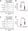
Stimulation with SEA induces the same biased ERK/p38 phosphorylation as anti-CD3. (A and C) Human CD4+ T cells were negatively enriched by magnetic-activated cell sorting and then stimulated with SEA-pulsed, autologous dendritic cells. The CD4+Vβ1+-cell population (unfract.) was gated, and phosphorylation of ERK (ppERK, A) or p38 (ppp38, C) was analyzed by Phosphoflow. (A) Unfractionated CD4+Vβ1+ cells stimulated for 4 min were divided into naïve (red) and experienced (blue) populations. (B) Phospho-ERK–expressing cell percentages of naïve and experienced populations were compared. Stimulation time was 4 min. (D) The same comparison as in B was conducted on phospho-p38. (B and D) Data represent the value of each experiment and the mean ± SD of eight independent experiments.
Fig. 4.
Fig. 4.
Distribution of phospho-SLP-76-expressing cells is skewed toward the CD4+ naïve T population. (A and B) SLP-76 phosphorylation (pSLP-76) in human PBMCs was analyzed by Phosphoflow. Cross-linking with 10 μg/mL anti-CD3ε (S4.1) and 5 μg/mL anti-CD28 (αCD3 + αCD28 crosslink) was used as a stimulant. (A) Cells were stimulated for 0, 1.5, 1.75, or 2 min, and CD4+ T-cell population (unfract. CD4+ population) was gated. (B) CD4+ T-cell population stimulated for 1.75 min was divided into naïve (red) and experienced (blue) populations. (C) SLP-76 phosphorylation levels of naïve and experienced populations were compared with normalized values. Values were calculated as in Fig. 1. Stimulation time was 1.75 min. Data represent the mean + SD of eight independent experiments.
Fig. 5.
Fig. 5.
hDlg associates with and phosphorylates p38 in CD4+ experienced cells but not in naïve cells upon TCR ligation. An immunoblot was used to analyze the capacity of hDlg to associate with and phosphorylate p38. (A and B) CD4+ experienced or naïve cells were negatively enriched by magnetic-activated cell sorting and stimulated with 10 μg/mL anti-CD3ε (S4.1) and 5 μg/mL anti-CD28 (αCD3 + αCD28 crosslink) for 2 min (B). Lysates were immunoprecipitated (IP) with anti-hDlg. Whole-cell lysate was also loaded (A). (C) Phosphorylation of p38 in naïve or experienced cells was quantitatively compared with the normalized values that were calculated as follows; the intensity of ppp38 band/that of the corresponding hDlg band. Data are representative of four independent experiments (A and B) and represent the mean + SD (C).
Fig. 6.
Fig. 6.
ERK phosphorylation induces an attenuated calcium influx and mediates the acceleration of signalsome degradation. (A and B) Human PBMCs were assayed for TCR-induced Ca2+ influx by flow cytometry. Cells were pretreated with DMSO, 10 μM U0126, or 20 μM PD98059 for 20 min. Arrow represents the time point for addition of precross-linked 10 μg/mL anti-CD3γ (UCHT1) and 5 μg/mL anti-CD28 (αCD3 + αCD28 crosslink) or the precross-linked isotype control IgG1 (iso. control). (B) CD4+ naïve (Upper) or experienced cells (Lower) were gated, and [Ca2+]i level in each population was analyzed. (C) Effect of ERK inhibitor on naïve and experienced populations was compared with the values calculated as follows: (peak Indo-1 ratio of ERK inhibitor-treated cells)/(that of DMSO-treated cells). Data represent the mean + SD of nine independent experiments. (D) Immunoblot was conducted to analyze the role of phospho-ERK in stability/instability of signalsome. CD4+ naïve cells were negatively enriched by magnetic-activated cell sorting, pretreated with DMSO or 10 μM U0126 for 20 min, and stimulated by cross-link with 10 μg/mL anti-CD3ε (S4.1) and 5 μg/mL anti-CD28 (αCD3 + αCD28 crosslink) for 0, 1.75, or 3.5 min. Lysates were immunoprecipitated (IP) with anti-LAT. Data are representative of three independent experiments.

References

    1. Smith-Garvin JE, Koretzky GA, Jordan MS. T cell activation. Annu Rev Immunol. 2009;27:591–619. - PMC - PubMed
    1. Wange RL. TCR signaling: another Abl-bodied kinase joins the cascade. Curr Biol. 2004;14:R562–R564. - PubMed
    1. van Oers NS. T cell receptor-mediated signs and signals governing T cell development. Semin Immunol. 1999;11:227–237. - PubMed
    1. Alberola-Ila J, Takaki S, Kerner JD, Perlmutter RM. Differential signaling by lymphocyte antigen receptors. Annu Rev Immunol. 1997;15:125–154. - PubMed
    1. Cantrell D. T cell antigen receptor signal transduction pathways. Annu Rev Immunol. 1996;14:259–274. - PubMed

Publication types

MeSH terms

LinkOut - more resources