Comparative Study
. 2011 Apr;337(1):110-6.
doi: 10.1124/jpet.110.176321. Epub 2010 Dec 30.

Acetaminophen-induced hepatotoxicity in mice occurs with inhibition of activity and nitration of mitochondrial manganese superoxide dismutase

Affiliations
Comparative Study

Acetaminophen-induced hepatotoxicity in mice occurs with inhibition of activity and nitration of mitochondrial manganese superoxide dismutase

Rakhee Agarwal et al. J Pharmacol Exp Ther. 2011 Apr.

Abstract

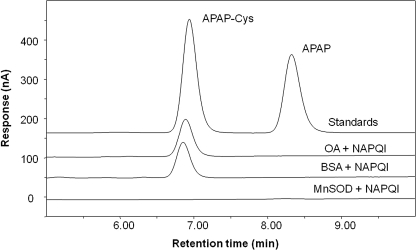
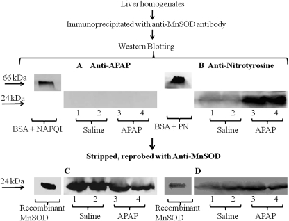
In overdose the analgesic/antipyretic acetaminophen (APAP) is hepatotoxic. Toxicity is mediated by initial hepatic metabolism to N-acetyl-p-benzoquinone imine (NAPQI). After low doses NAPQI is efficiently detoxified by GSH. However, in overdose GSH is depleted, NAPQI covalently binds to proteins as APAP adducts, and oxygen/nitrogen stress occurs. Toxicity is believed to occur by mitochondrial dysfunction. Manganese superoxide dismutase (MnSOD) inactivation by protein nitration has been reported to occur during other oxidant stress-mediated diseases. MnSOD is a critical mitochondrial antioxidant enzyme that prevents peroxynitrite formation within the mitochondria. To examine the role of MnSOD in APAP toxicity, mice were treated with 300 mg/kg APAP. GSH was significantly reduced by 65% at 0.5 h and remained reduced from 1 to 4 h. Serum alanine aminotransferase did not significantly increase until 4 h and was 2290 IU/liter at 6 h. MnSOD activity was significantly reduced by 50% at 1 and 2 h. At 1 h, GSH was significantly depleted by 62 and 80% at nontoxic doses of 50 and 100 mg/kg, respectively. No further GSH depletion occurred with hepatotoxic doses of 200 and 300 mg/kg APAP. A dose response decrease in MnSOD activity was observed for APAP at 100, 200, and 300 mg/kg. Immunoprecipitation of MnSOD from livers of APAP-treated mice followed by Western blot analysis revealed nitrated MnSOD. APAP-MnSOD adducts were not detected. Treatment of recombinant MnSOD with NAPQI did not produce APAP protein adducts. The data indicate that MnSOD inactivation by nitration is an early event in APAP-induced hepatic toxicity.


Figures

Fig. 1.
Fig. 1.
Time course for effect of APAP on serum ALT, hepatic GSH, hepatic MnSOD activity, and hepatic MnSOD protein. Mice (n = 3) were treated with a 300 mg/kg dose of APAP and sacrificed at the indicated times, and serum and liver were collected. The 0 time is the saline-treated control mice. A, ALT levels in serum (hepatotoxicity). B, GSH levels in liver. C, MnSOD activity in liver. D, MnSOD protein levels in liver. Western blot analyses were performed on each homogenate using anti-MnSOD. GAPDH was used as a loading control. The data are presented as mean ± S.E. of the relative intensity. One representative gel is shown above the density values. *, significant difference from saline, p ≤ 0.05.
Fig. 2.
Fig. 2.
Dose response for effect of APAP on serum ALT, hepatic GSH, hepatic MnSOD activity, and hepatic MnSOD protein at 1 h. Mice (n = 3) were treated with 0, 50, 100, 200, and 300 mg/kg APAP and sacrificed after 1 h. A, ALT levels in serum (hepatotoxicity). B, GSH levels in liver. C, MnSOD activity in liver. D, MnSOD protein levels in liver. Western blot analyses were performed on each homogenate using anti-MnSOD. GAPDH was used as a loading control. Data are presented as mean ± S.E. of the relative intensity. One representative gel is shown above the density values. *, significant difference from saline, p ≤ 0.05.
Fig. 3.
Fig. 3.
HPLC-EC analysis of NAPQI-treated MnSOD, BSA, and OVA for APAP-Cys. BSA, OA, and recombinant MnSOD were reacted with synthetic NAPQI. The resultant proteins were gel-filtered, treated with protease, and analyzed by HPLC/EC. HPLC reference standards were APAP-Cys (6.85 min, 20 pmol on column) and APAP (8.33 min, 13.2 pmol on column). Detection signal is current in nA at a potential of 180 mV versus a palladium reference electrode.
Fig. 4.
Fig. 4.
Western blot of immunoprecipitated MnSOD for APAP covalent binding and tyrosine nitration. Liver homogenates were immunoprecipitated with anti-MnSOD antibody and resolved in 10% SDS-PAGE followed by Western blot analysis. Lanes 1 and 2 are from saline-treated mouse livers. Lanes 3 and 4 are from APAP-treated (300 mg/kg) mouse liver at 1 h. A and B, Western blot analyses were performed using anti-APAP antibody (A) and anti-3-nitrotyrosine antibody (B). C and D, subsequently, the blots were stripped and reprobed with anti-MnSOD antibody. BSA treated with NAPQI (A), peroxynitrite (B) at 66 kDa, and recombinant MnSOD at 24 kDa (C and D) were used as positive controls. One representative experiment is shown.
Fig. 5.
Fig. 5.
APAP time course and dose response for nitration of MnSOD. A, the samples from the time course (Fig. 1D) were stripped and reprobed with anti-3-nitrotyrosine antibody. B, the samples from the dose response (Fig. 2D) were stripped and reprobed with anti-3-nitrotyrosine antibody. The data are presented as mean ± S.E. of the relative intensity. One representative gel is shown. *, significant difference from saline, p ≤ 0.05.
Fig. 6.
Fig. 6.
Schematic representation of how reactive nitrogen can alter oxidant stress in mitochondria.

References

    1. Bayir H, Kagan VE, Clark RS, Janesko-Feldman K, Rafikov R, Huang Z, Zhang X, Vagni V, Billiar TR, Kochanek PM. (2007) Neuronal NOS-mediated nitration and inactivation of manganese superoxide dismutase in brain after experimental and human brain injury. J Neurochem 101:168–181 - PubMed
    1. Beckman JS. (1996) Oxidative damage and tyrosine nitration from peroxynitrite. Chem Res Toxicol 9:836–844 - PubMed
    1. Boobis AR, Tee LB, Hampden CE, Davies DS. (1986) Freshly isolated hepatocytes as a model for studying the toxicity of paracetamol. Food Chem Toxicol 24:731–736 - PubMed
    1. Burcham PC, Harman AW. (1991) Acetaminophen toxicity results in site-specific mitochondrial damage in isolated mouse hepatocytes. J Biol Chem 266:5049–5054 - PubMed
    1. Burke AS, MacMillan-Crow LA, Hinson JA. (2010) Reactive nitrogen species in acetaminophen-induced mitochondrial damage and toxicity in mouse hepatocytes. Chem Res Toxicol 23:1286–1292 - PMC - PubMed

Publication types

MeSH terms