. 2011 Feb;121(2):554-68.
doi: 10.1172/JCI43984. Epub 2011 Jan 4.

Vaccine-induced protection against 3 systemic mycoses endemic to North America requires Th17 cells in mice

Affiliations

Vaccine-induced protection against 3 systemic mycoses endemic to North America requires Th17 cells in mice

Marcel Wüthrich et al. J Clin Invest. 2011 Feb.

Erratum in

Abstract

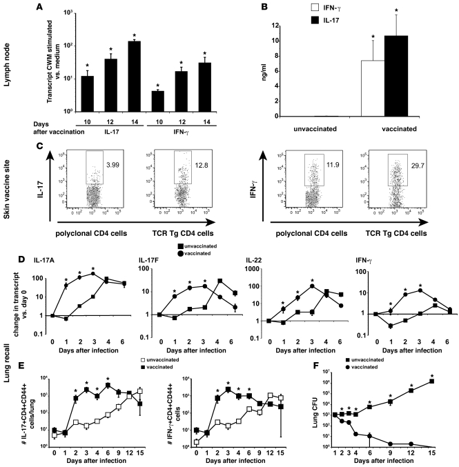
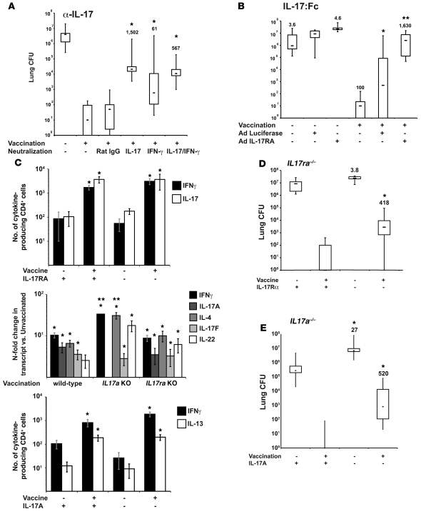
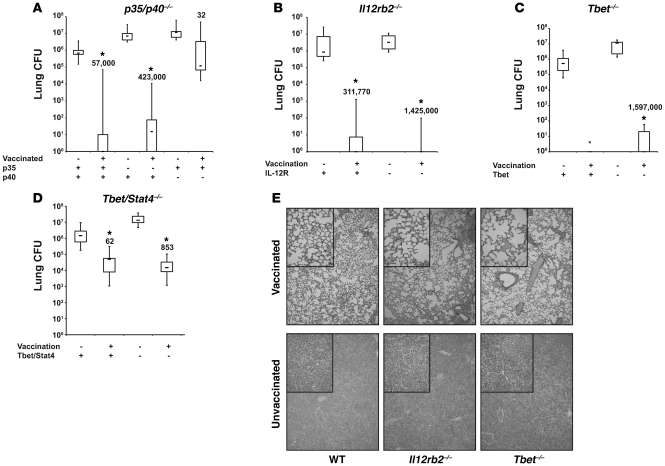
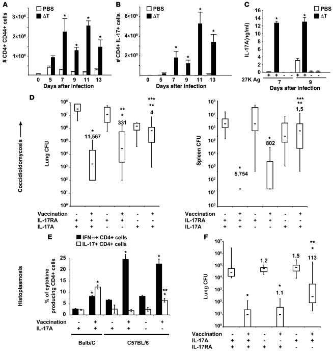
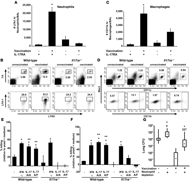
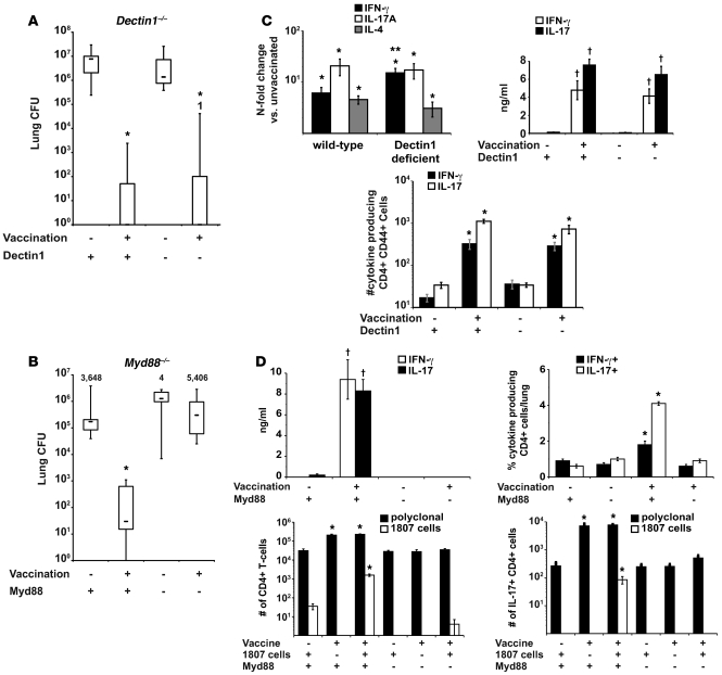
Worldwide rates of systemic fungal infections, including three of the major pathogens responsible for such infections in North America (Coccidioides posadasii, Histoplasma capsulatum, and Blastomyces dermatitidis), have soared recently, spurring interest in developing vaccines. The development of Th1 cells is believed to be crucial for protective immunity against pathogenic fungi, whereas the role of Th17 cells is vigorously debated. In models of primary fungal infection, some studies have shown that Th17 cells mediate resistance, while others have shown that they promote disease pathology. Here, we have shown that Th1 immunity is dispensable and that fungus-specific Th17 cells are sufficient for vaccine-induced protection against lethal pulmonary infection with B. dermatitidis in mice. Further, vaccine-induced Th17 cells were necessary and sufficient to protect against the three major systemic mycoses in North America. Mechanistically, Th17 cells engendered protection by recruiting and activating neutrophils and macrophages to the alveolar space, while the induction of Th17 cells and acquisition of vaccine immunity unexpectedly required the adapter molecule Myd88 but not the fungal pathogen recognition receptor Dectin-1. These data suggest that human vaccines against systemic fungal infections should be designed to induce Th17 cells if they are to be effective.


Figures

Figure 1
Figure 1. Naive CD4 T cells differentiate into Th17 cells during the afferent phase of vaccination and are recalled to the lung during the efferent phase.
(A) Ag-specific Th17 cells accumulate in the skin-draining lymph nodes during vaccine induction. Purified CD4 T cells from the skin-draining lymph nodes of vaccinated mice were cocultured with CW/M Ag for 2 days and cytokine transcripts analyzed by RT-PCR. Mean ± SEM (n = 3); representative of 3 experiments. (B) Primed Th17 cells produce IL-17 in response to in vitro Ag stimulation. 3 weeks after the boost, purified CD4 T cells from the draining nodes were stimulated with CW/M Ag for 3 days and the supernatants analyzed. Mean ± SEM (n = 4); representative of 3 experiments. (C) Th17 cells infiltrate the vaccine site. Lymphocytes were isolated from the vaccine site. To verify cytokine response by Ag-specific T cells, B. dermatitidis–specific 1807 Tg cells were transferred into recipients before vaccination. T cells from the site were stimulated with α-CD3 and α-CD28 mAb for 4 hours and analyzed for intracellular cytokine. Dot plots show percentage of cytokine-producing polyclonal or Tg CD4 cells. (D and E) Th17 effector cells migrate to the lung during the recall and vaccine efferent phase. Vaccinated mice were challenged with B. dermatitidis i.t. and the expression of lung cytokine transcript was measured by RT-PCR. For the kinetics of IL-17– and IFN-γ– producing CD4 T cells, the number of cytokine-producing cells was calculated by multiplying the number of total lung cells by the percentage of cytokine-producing CD4 T cells. Mean ± SEM (n = 4); representative of 3 experiments. (F) Lung CFUs coincide with number of cytokine-producing CD4 T cells. Mean ± SEM (n = 8–10); representative of 2 experiments. *P < 0.05 versus unvaccinated mice.
Figure 2
Figure 2. IL-17 mediates vaccine immunity against B. dermatitidis infection.
(A) Antibody neutralization of IL-17 in vaccinated mice after infection. Lung CFU are the mean ± SEM (n = 10–12). Numbers shown are the relative increase in lung CFU of mAb-treated versus rat IgG controls. *P < 0.001 versus vaccinated mice treated with rat IgG. (B) Neutralization of IL-17 by soluble IL-17 receptor (IL-17R:Fc). Values are the mean ± SEM (n = 10–18); representative of 2 experiments. Numbers shown are the fold increase in lung CFU versus AdLuc-treated controls. *P < 0.001 versus vaccinated mice not treated with adenovirus; **P < 0.001 versus vaccinated mice treated with AdLuc. (C) Lung transcripts and number of primed Th1 and Th17 cells recruited to the lung upon fungal challenge in Il17a–/– and Il17ra–/– mice. RNA from lung (middle panel) and the absolute number of IL-17–, IFN-γ–, or IL-13–producing CD4+CD44+ T cells were quantified by real-time PCR and FACS. Data are mean ± SEM (n = 4–6); representative of 2 experiments. *P < 0.05 versus unvaccinated controls; **P < 0.05 versus vaccinated wild-type and Il17ra–/– mice (D) IL-17RA is required for vaccine immunity. Mean ± SEM (n = 8–12); representative of 2 experiments. *P < 0.001 versus vaccinated wild-type. (E) IL-17A is required for vaccine immunity. Lung CFU are the mean ± SEM (n = 10–12); representative of 2 experiments. Numbers shown in D and E are the fold increase in CFU versus corresponding wild-type controls. *P < 0.001 versus wild-type mice.
Figure 3
Figure 3. Th1 cells are dispensable in vaccine immunity to fungi.
(A) Il12p35–/– and Il12/23p40–/–, (B) Il12rb2–/–, (C) Tbet–/–, or (D) Tbet–/–Stat4–/– and wild-type mice were vaccinated and challenged with B. dermatitidis as in Methods and the lung CFU analyzed. CFU values are the mean ± SEM of 10–12 mice/group; representative of 2 experiments per group. The numbers shown are fold reduction in lung CFU versus unvaccinated controls. *P < 0.001 versus unvaccinated controls and vaccinated p40–/– mice. (E) Vaccine-induced Th17 cells do not mediate lung pathology. Histology (16 days after infection) of lungs from wild-type and Th1-deficient vaccinated mice showed normal lung alveolar parenchyma with few foci of perivascular lymphocytes, but no PMN infiltrates. In contrast, lungs from unvaccinated mice showed massive infiltration with PMNs and macrophages and many yeast. Original magnification, ×100; ×400 (insets).
Figure 4
Figure 4. Ag-specific Th17 cells are sufficient to mediate antifungal resistance.
(A) Polarization of Th17 cells. The dot plot shows the percentage of IL-17– and IFN-γ–positive CD4+ cells and is representative of all 4 types of TCR Tg T cells (below). (B and C) Transfer of Th17-polarized cells into wild-type recipients. Th17-polarized cells from wild-type × 1807, Il12rb2–/– × 1807, Tbet–/– × 1807, and OT2 mice were transferred into nonirradiated or sublethally irradiated wild-type mice (78). The next day (nonirradiated recipients) or 8 weeks later (irradiated recipients), mice were challenged with B. dermatitidis and analyzed for lung CFU 2 weeks later. Values are the mean ± SEM (n = 11–18). Numbers shown are fold reduction in lung CFU versus OT2 controls. *P < 0.05 versus OT2 controls. (D) In vivo primed Th17 cells protect vaccinated OT1 mice. 106 wild-type × 1807, Il12rb2–/– × 1807, and Tbet–/– × 1807 cells were transferred into OT1 mice. Recipients were vaccinated, challenged, and analyzed for lung CFU. Values are the mean ± SEM (n = 9–12). Numbers shown are the fold reduction in lung CFU versus OT2 controls. *P < 0.05 versus unvaccinated mice and vaccinated mice that received OT2 cells. (E) T helper phenotype of in vivo primed Il12rb2–/– × 1807 and Tbet–/– × 1807 cells. At day 4 after infection, the percentages of cytokine-producing transferred Tg cells were quantified (4–6 mice per group). *P < 0.05 versus 1807 × Tbet–/– and 1807 × Il12rb2–/– cells. (F) IL-17 and IFN-γ production by splenocytes in response to CW/M Ag in vitro was analyzed; *P < 0.05 versus all other groups.
Figure 5
Figure 5. Th17 cells mediate vaccine immunity to multiple dimorphic fungi.
(AD) C. posadasii. C57BL/6 mice were vaccinated with the live attenuated strain (ΔT) and challenged with C. posadasii spores as in Methods. The phenotype of lung CD4 T cells was analyzed serially after infection (A and B). IL-17 production by splenocytes in response to T27K Ag was analyzed in vitro as in Methods (C); *P < 0.05 versus respective PBS control (AC). Vaccinated mice were challenged and lung CFU analyzed 2 weeks after infection (D). The numbers shown are fold reduction in CFU versus respective control. *P < 0.001 versus unvaccinated controls; **P < 0.05 versus vaccinated wild-type mice; ***P < 0.05 versus vaccinated Il17a–/– mice. (E) H. capsulatum. Balb/C and C57BL/6 mice were vaccinated s.c. and challenged with a sublethal dose of H. capsulatum yeast as in Methods. At day 4 after infection, the percentages of IL-17– and IFN-γ–producing CD4+ T cells were assayed by intracellular cytokine staining and FACS. Data are the mean ± SEM of 4 mice/group. *P < 0.01 versus unvaccinated mice; **P < 0.01 versus vaccinated wild-type mice. (F) C57BL/6 wild-type, Il17ra–/–, and Il17a–/– mice were vaccinated and challenged as in Methods. At day 14 after infection, the mice were sacrificed and analyzed for the burden of lung infection. Data are the mean ± SEM of 10 to 20 mice per group. *P < 0.01 versus unvaccinated mice; **P < 0.01 versus vaccinated wild-type mice.
Figure 6
Figure 6. Th17 cells are instrumental in phagocyte recruitment and activation.
Il17ra–/– and wild-type mice were vaccinated with B. dermatitidis and challenged. At day 4 after infection, cells from BAL fluid were harvested, enumerated, and stained by FACS. (A) The number of LFA1+ PMNs in BAL; mean ± SEM of 4–10 mice/group. *P < 0.05 versus unvaccinated wild-type and vaccinated Il17ra–/– mice. (B) The percentage of LFA1+ PMNs (CD11b+, 7/4+, and Ly6G+) in BAL; mean ± SEM (n = 4). (C) The number of CD11b+ macrophages (CD11c+, Mac3+) in BAL; mean ± SEM (n = 4). *P < 0.05 versus unvaccinated wild-type and vaccinated Il17ra–/– mice. (D) The percentage of CD11b+ macrophages (CD11c+, Mac3+) in BAL; mean ± SEM (n = 4). (E and F) IL-17–induced killing by neutrophils and alveolar macrophages. Neutrophils and alveolar macrophages from naive wild-type and Il17ra–/– mice were activated with 200 ng/ml of IFN-γ, homodimeric IL-17A/A, or heterodimeric IL-17A/F. B. dermatitidis yeast were added to cells at a MOI of 0.01 and cocultured for 4 hours with neutrophils and 24 hours with macrophages. Cell culture supernatants and cell lysates were combined and cultured to enumerate CFU. *P < 0.05 versus resting neutrophils and macrophages; **P < 0.05 versus corresponding cocultures with cells from Il17ra–/– mice. (G) Influence of neutrophil depletion on vaccine immunity. Neutrophils in vaccinated and unvaccinated wild-type mice were depleted with 250 μg of 1A8 mAb injected i.v. at days 0 and 2 after infection. As a control, mice received rat IgG. At day 4 after infection, lung CFU were enumerated. Data represent an average ± SEM (n = 10–12). *P < 0.001 versus corresponding rat IgG–treated mice. The numbers shown are the fold increase in CFU in neutrophil-depleted versus nondepleted mice.
Figure 7
Figure 7. Vaccine-induced resistance is independent of Dectin-1, but dependent on Myd88.
(A) Dectin1–/– mice and wild-type controls were vaccinated with B. dermatitidis. 2 weeks after challenge, mice were analyzed for lung CFU. *P < 0.001 versus unvaccinated control mice. (B) Myd88–/– mice and wild-type mice were vaccinated with 106 heat-killed yeast. Mice were challenged and 2 weeks later, when the unvaccinated controls were moribund, lung CFU were enumerated. Data are the mean ± SEM (n = 8–12). *P < 0.001 versus all other groups. (C and D) Lung transcript was analyzed 2 days after infection. RNA was isolated from lung, and transcripts were analyzed by RT-PCR. Data are expressed as fold change versus unvaccinated mice and represent the mean ± SEM (n = 4–6). *P < 0.05 versus transcript in unvaccinated controls; **P < 0.05 versus transcript in vaccinated wild-type mice. Intracellular cytokine staining was done at day 3–4 after infection. Lung cells were stained and the absolute numbers of cytokine-producing CD4+ CD44+ T cells determined by FACS. Data represent the mean ± SEM (n = 4). *P < 0.05 versus intracellular cytokine in unvaccinated wild-type controls and vaccinated Myd88–/– mice (C and D). To assay Ag-specific cytokine production, CD4+ cells were purified from the skin-draining lymph nodes and spleen and stimulated with CW/M Ag for 2 days. Supernatant was assayed by ELISΑ. Data are the mean ± SEM of 4 independent experiments (n = 4 mice/group). P < 0.001 versus cytokine production by CD4 cells from unvaccinated controls or vaccinated Myd88–/– mice (C and D).

Comment in

Similar articles

Cited by

References

    1. Pulendran B, Seder RA. Host-pathogen interactions in the 21st century. Curr Opin Immunol. 2005;17(4):335–337. doi: 10.1016/j.coi.2005.06.004. - DOI - PMC - PubMed
    1. Cutler JE, Deepe GS, Jr, Klein BS. Advances in combating fungal diseases: vaccines on the threshold. Nat Rev Microbiol. 2007;5(1):13–28. doi: 10.1038/nrmicro1537. - DOI - PMC - PubMed
    1. Mochon AB, Cutler JE. Is a vaccine needed against Candida albicans? Med Mycol. 2005;43(2):97–115. doi: 10.1080/13693780500035979. - DOI - PubMed
    1. Deepe GS, Jr, Wuthrich M, Klein BS. Progress in vaccination for histoplasmosis and blastomycosis: coping with cellular immunity. Med Mycol. 2005;43(5):381–389. doi: 10.1080/13693780500245875. - DOI - PubMed
    1. Cole GT, et al. A vaccine against coccidioidomycosis is justified and attainable. Med Mycol. 2004;42(3):189–216. doi: 10.1080/13693780410001687349. - DOI - PubMed

Publication types

MeSH terms